Sina Amirrajab1, William Paul Segars2, Cristian Lorenz3, Juergen Weese3, and Marcel Breeuwer1,4
1Biomedical Engineering Department, Eindhoven University of Technology, Eindhoven, Netherlands, 2Carl E. Ravin Advanced Imaging Laboratories, Duke University, Durham, NC, United States, 3Philips Research Laboratories, Hamburg, Germany, 4MR R&D - Clinical Science, Philips Healthcare, Best, Netherlands
1Biomedical Engineering Department, Eindhoven University of Technology, Eindhoven, Netherlands, 2Carl E. Ravin Advanced Imaging Laboratories, Duke University, Durham, NC, United States, 3Philips Research Laboratories, Hamburg, Germany, 4MR R&D - Clinical Science, Philips Healthcare, Best, Netherlands
Based upon the high-resolution ex-vivo data we demonstrated
that accurate modelling and Inclusion of detail anatomy of the endocardial
trabeculae into the XCAT heart model provides a greater realism for cardiac MR
image simulation.
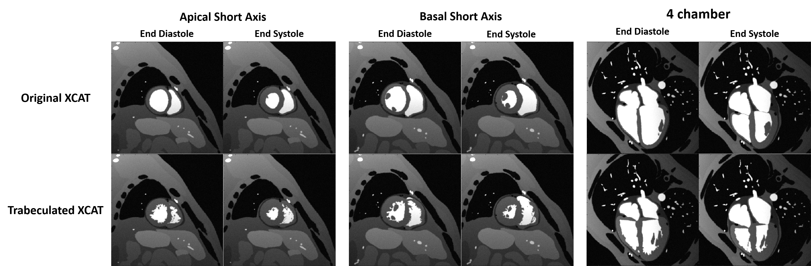
Figure 3 Simulation of MR images. The short
axis view at apical and basal slices and 4-chamber view of the original XCAT heart
(top row) and the same views for trabeculated XCAT (bottom row) were simulated at
end diastolic and end systolic of heart.
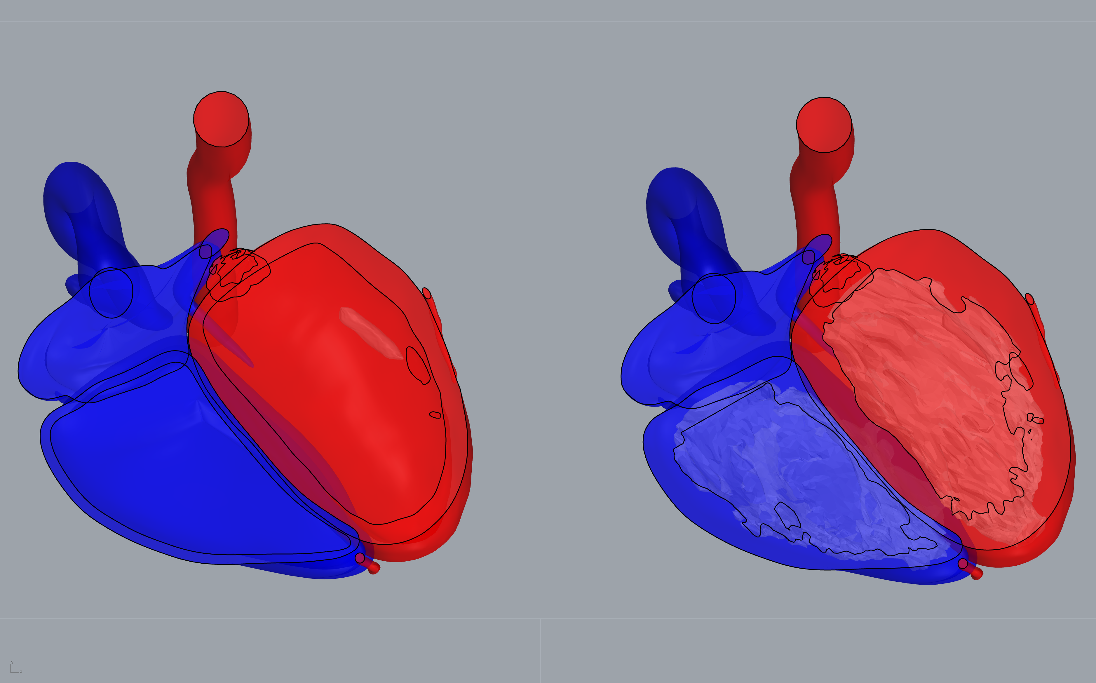
Figure 2 Alignment of the trabeculae model of
the LV (red) and the RV (blue) within the inner layer of the original XCAT
heart using Rhinoceros surface modelling software. The procedure involves 3D
nonlinear transformation and rotation of the trabeculae model to have it
incorporated into the LV and RV surfaces.
