Ruponti Nath1, Sean Callahan1, Narayana Singam2, Marcus Stoddard2, and Amir Amini1
1ECE, University of Louisville, Louisville, KY, United States, 2Cardiovascular Medicine, University of Louisville, Louisville, KY, United States
1ECE, University of Louisville, Louisville, KY, United States, 2Cardiovascular Medicine, University of Louisville, Louisville, KY, United States
Reconstruction of 2D phase contrast MRI from undersampled K space by deep convolutional neural networks is proposed.

Figure 1: Proposed
U-net architecture. It has 7 convolution layer in contraction and 7 convolution
layer in expansion. After each two convolution layer in contraction there is a
maximum pooling layer. In expansion average unpooling is done to restore image
resolution. Black, red and blue arrow denotes convolution , pooling and
unpooling operation. Green arrow shows concatenation from contraction layer to
expansion layer.
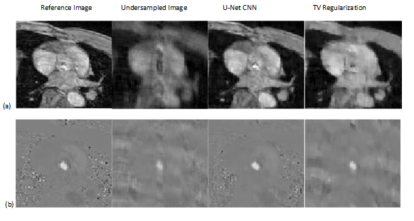
Figure 2. (a)
shows magnitude image and Figure 2(b)
shows Phase Image from reference complex image, undersampled complex image, image reconstructed by proposed U-net and image reconstructed by TV regularization. The image is in the peak systole phase of the
cardiac cycle and is exactly at the location of the aortic valve.