Edward M Lawrence1, Yuxin Zhang1, Jitka Starekova1, Zihan Wang2, Shane A Wells1, and Diego Hernando1
1Radiology, University of Wisconsin-Madison, Madison, WI, United States, 2Biomedical Engineering, University of Wisconsin-Madison, Madison, WI, United States
1Radiology, University of Wisconsin-Madison, Madison, WI, United States, 2Biomedical Engineering, University of Wisconsin-Madison, Madison, WI, United States
In
a prospective clinical cohort, reduced-distortion DWI methods, based on reduced
field of view and multi-shot techniques, enable improved image quality and
reproducible ADC quantification in prostate imaging, compared to standard single-shot
EPI.
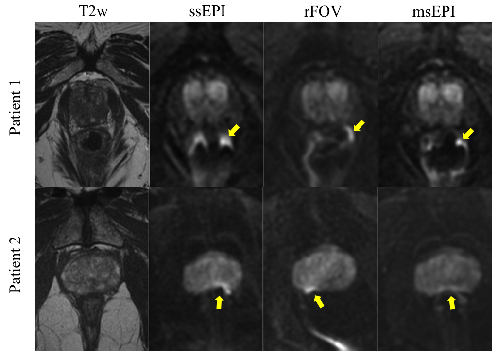
Figure 1. Representative images from two patients demonstrating reduced artifactual distortion on DWI with rFOV and msEPI. The qualitative improvement in distortion is greatest for msEPI (right column) in both patients which results in reduced signal buildup due to rectal gas (yellow arrows) and less anterior-posterior distortion of the prostate shape, especially for patient 2 (ssEPI, single shot echo planar image; rFOV, reduced field of view; msEPI, multi-shot echo planar image)
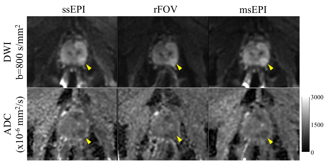
Figure 2. 60-yo male with elevated prostate specific antigen (4.46 ng/mL) and suspicious lesion on prostate MRI. The suspicious lesion (yellow arrows) is seen in the left peripheral zone on all DWI sequences, including b=800 series (top row) and calculated ADC map (bottom row, ADC color bar on right). Mean ADC for the lesion was 1075.8, 1098.8, and 1067.5 x10-6mm2/s on ssEPI, rFOV, and msEPI, respectively. The patient subsequently was found to have Gleason 3+4 disease at surgery. (ssEPI, single shot echo planar image; rFOV, reduced field of view; msEPI, multi-shot echo planar image)