1Department of Radiology, University of Wisconsin-Madison, Madison, WI, United States, 2Medical Physics, University of Wisconsin-Madison, Madison, WI, United States
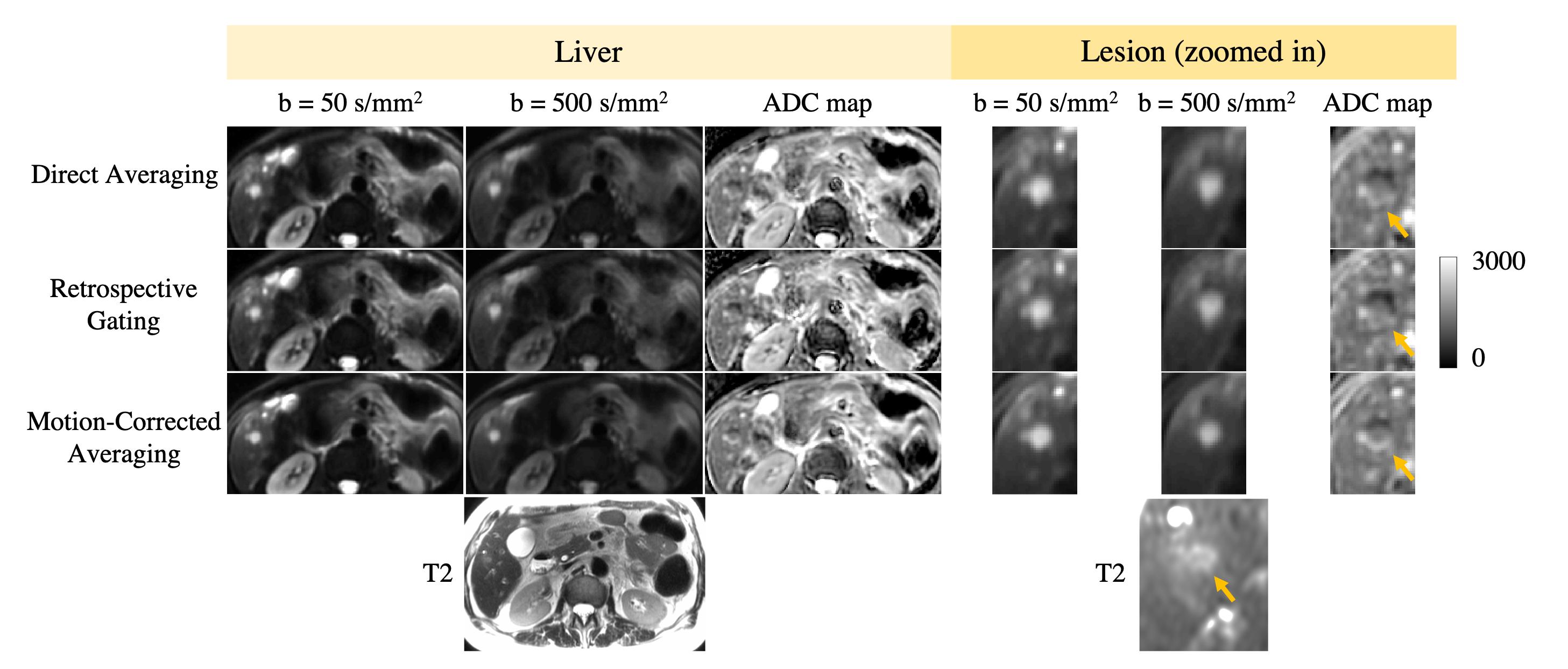
Fig.2. DW images and ADC maps of a liver metastatic lesion from pancreatic cancer, processed with direct averaging, retrospective gating, and motion-corrected averaging (NLM). The lesion appeared extended in size with blurry boundaries in DW images and ADC maps processed with direct averaging and retrospective gating, due to inadequate alignment of the lesion across repetitions and b values. And retrospective gating had lower SNR. With NLM, shape and size of the lesion showed improved correspondence to co-localized T2 reference, and boundaries appeared clearer in DWI and ADC map.
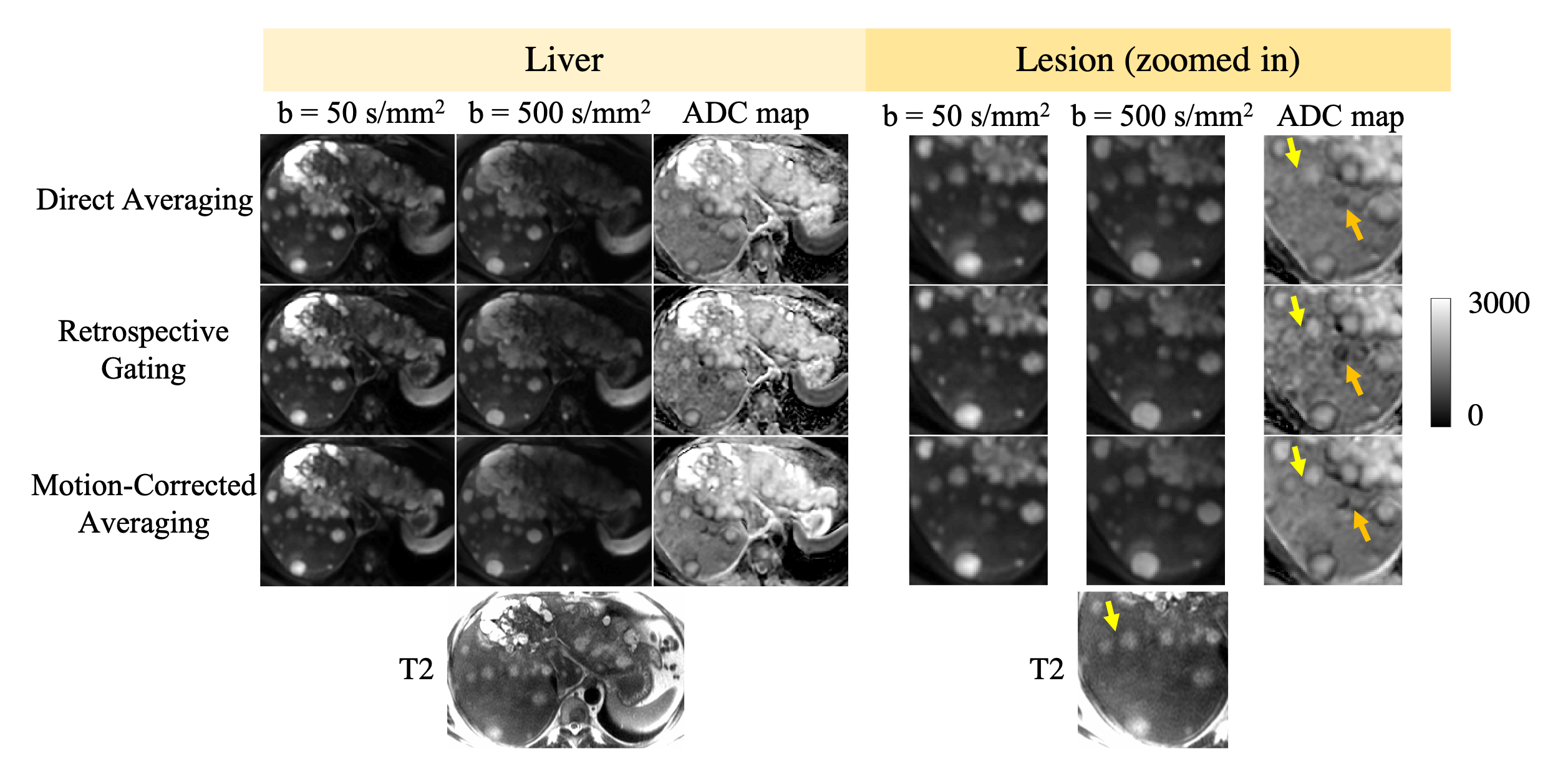
Fig.3. DW images and ADC maps of multiple liver metastatic lesions from pancreatic cancer processed with direct averaging, retrospective gating, and motion-corrected averaging (NLM). Two lesions (see yellow arrows) had blurry boundaries in DW images and ADC maps processed with direct averaging and retrospective gating. With NLM, boundaries were clearly delineated and showed improved correspondence to co-localized T2 reference. In this challenging case, NLM did not align every lesion correctly across b-values, as in a black crescent due to mis-registration (see orange arrows).