Hui Wang1,2,3, Amol Pednekar2,3, Jean A. Tkach2,3, Charles L. Dumoulin2,3, Kaley R. Bridgewater2, Andrew T. Trout2,3, and Jonathan R. Dillman2,3
1Philips, Cincinnati, OH, United States, 2Department of Radiology, Cincinnati Children’s Hospital Medical Center, Cincinnati, OH, United States, 3Department of Radiology, University of Cincinnati College of Medicine, Cincinnati, OH, United States
1Philips, Cincinnati, OH, United States, 2Department of Radiology, Cincinnati Children’s Hospital Medical Center, Cincinnati, OH, United States, 3Department of Radiology, University of Cincinnati College of Medicine, Cincinnati, OH, United States
We describe a 3D fast field echo (FFE) Magnetic
Resonance Elastography (MRE) pulse sequence for measurement of liver stiffness
in a single breath hold.
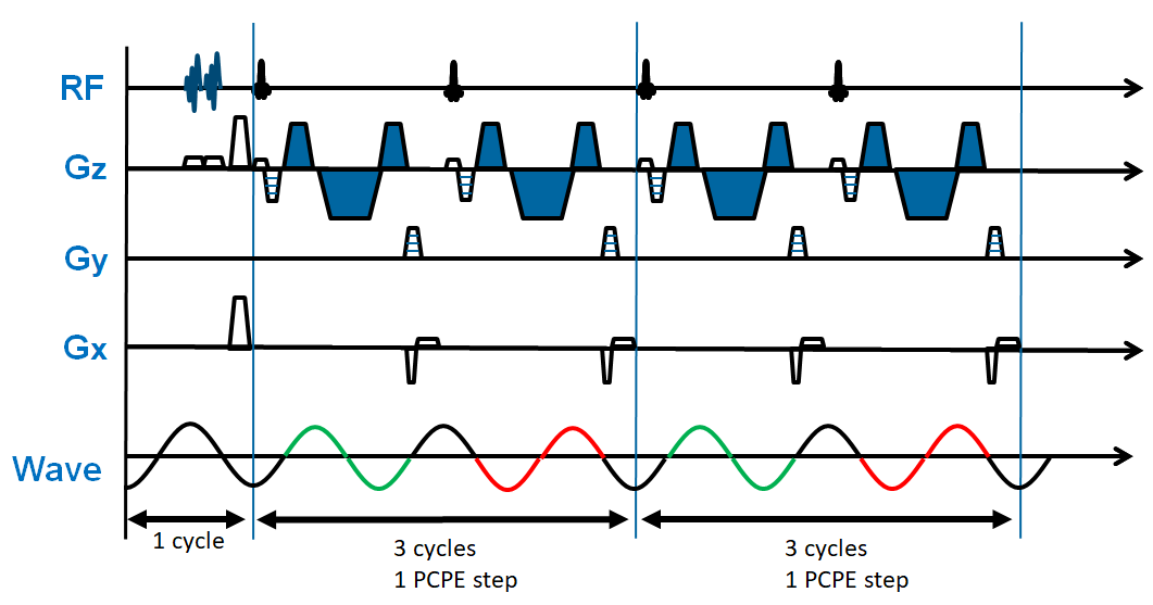
Figure 1. Schematic diagram of proposed
fast 3D MRE pulse sequence. Flow suppression pre-pulse are applied within one motion
cycle (16.67ms at 60Hz frequency) and are interleaved with variable phase-contrast
phase-encoding (PCPE) steps. In this diagram, two PCPE steps are shown. Each
PCPE step consists of two RF excitations with repetition time of 1.5 motion
cycles. The polarity of MEGs remains the same across TRs, as illustrated by the
MEG denoted in blue. Gz = slice select direction, Gy = phase encoding direction,
Gx = readout direction.
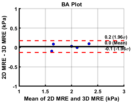
Figure 3. Bland-Altman Plot of conventional 2D MRE versus fast 3D MRE
derived stiffness values in 5 volunteers. Center line shows no mean difference
in stiffness values between these two techniques, and the upper/lower dotted
lines demarcate the 95% limits of agreement.