Cong Sun1, Yufan Chen1, Jinxia Zhu2, and Guangbin Wang1
1Shandong Medical Imaging Research Institute, Shandong University, Jinan, China, 2MR Collaboration, Healthcare Siemens Ltd., Beijing, China, Beijing, China
1Shandong Medical Imaging Research Institute, Shandong University, Jinan, China, 2MR Collaboration, Healthcare Siemens Ltd., Beijing, China, Beijing, China
MRI was significantly
better than ultrasound in being able to diagnose normal and abnormal fetal CC.
Also, fetal MRI can be helpful in assessing associated abnormalities and enhancing
prognostic consultations.
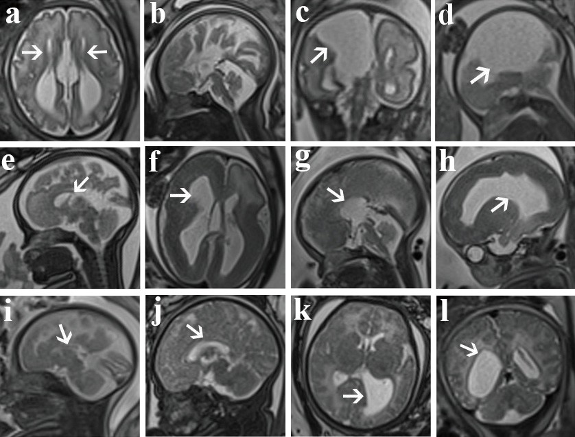
Fig. 2. a-b, A fetus
at 33 weeks of gestation with isolated complete absence of the corpus callosum
(CACC). c-d A fetus at 30 weeks of gestation. CACC with a huge
intra-hemispheric cyst. e, A fetus at 32 weeks of gestation with isolated splenium
absence (PACC). f-h A fetus at 33 weeks of gestation. A partial absence of the CC
(PACC) with pachygyria, and left front lobe leukodystrophy. i, A fetus at 28
weeks of gestation with isolated hypogenesis (HCC) and the CC is significantly
thinner and shorter. J-l A fetus at 35 weeks of gestation. HCC with bilateral
frontal lobe leukodystrophy.
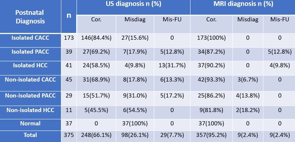
Table 3. Diagnosing fetal CC abnormalities in the postnatal, ultrasound (US), and magnetic resonance imaging (MRI) diagnostic groups
Cor., correct diagnosis; Misdiag, misdiagnosis; Mis-FU, missed diagnosis Isolated CACC, Isolated complete agenesis of the corpus callosum; Isolated PACC, Isolated partial agenesis of the corpus callosum; Isolated HCC, Isolated hypoplasia of the corpus callosum; Non-isolated CACC, CACC with other malformations; Non-isolated PACC: PACC with other malformations; Non-isolated HCC: HCC with other malformations.