Qiang Liu1, Zhongbiao Xu2, Xinyuan Zhang1, Zhifeng Chen1, Rongli Zhang3, Yaohui Wang4, Kaixuan Zhao1, Ed X. Wu5,6, and Yanqiu Feng1
1School of Biomedical Engineering, Guangdong Provincial Key Laboratory of Medical Image Processing, Southern Medical University, Guangzhou, China, 2Department of Radiotherapy, Cancer Center, Guangdong Provincial People's Hospital & Guangdong Academy of Medical Science, Guangzhou, China, 3School of Medicine, Guangdong Provincial People's Hospital , South China University of Technology, Guangzhou, China, 4Institute of Electrical Engineering, Chinese Academy of Sciences, Beijing, China, 5Laboratory of Biomedical Imaging and Signal Processing, The University of Hong Kong, Hong Kong SAR, China, 6Department of Electrical and Electronic Engineering, The University of Hong Kong, Hong Kong SAR, China
1School of Biomedical Engineering, Guangdong Provincial Key Laboratory of Medical Image Processing, Southern Medical University, Guangzhou, China, 2Department of Radiotherapy, Cancer Center, Guangdong Provincial People's Hospital & Guangdong Academy of Medical Science, Guangzhou, China, 3School of Medicine, Guangdong Provincial People's Hospital , South China University of Technology, Guangzhou, China, 4Institute of Electrical Engineering, Chinese Academy of Sciences, Beijing, China, 5Laboratory of Biomedical Imaging and Signal Processing, The University of Hong Kong, Hong Kong SAR, China, 6Department of Electrical and Electronic Engineering, The University of Hong Kong, Hong Kong SAR, China
This work aimed to
improve rat kidney diffusion-weighted imaging (DWI) by using interleaved multishot
echo planar imaging (EPI) with 2D navigators. The in vivo rat imaging resulted in improved resolution and less geometric distortion.
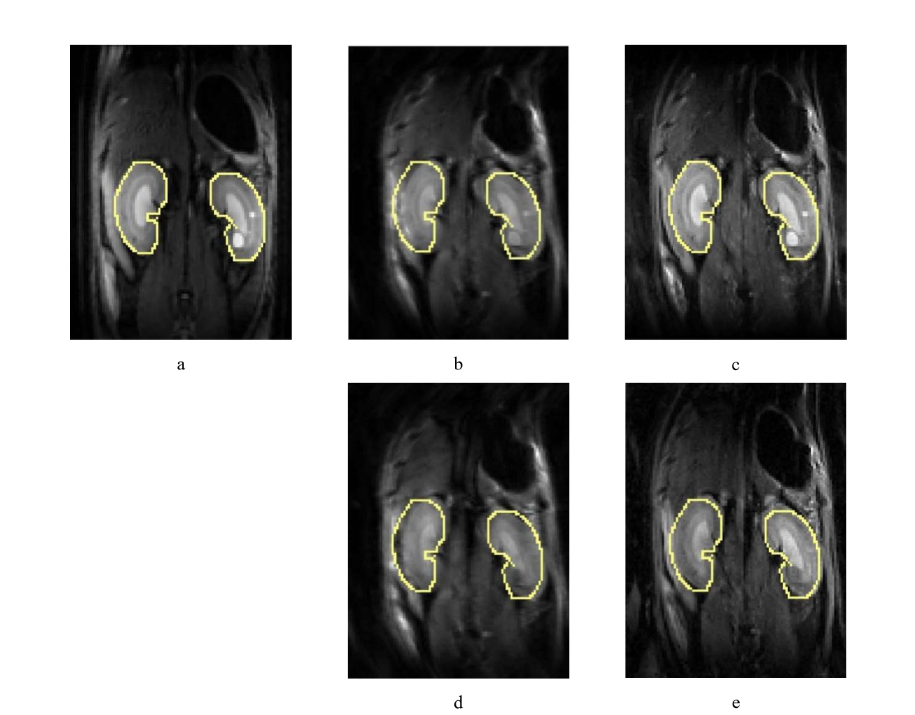
Figure
1 (a) T2-weighted image as reference. (b) ss-EPI for non-DW. (c)
2D navigated ms-EPI for non-DW.(d) DW acquisition with b value= 500 s/mm2 for ss-EPI
. (e) DW acquisition with b value= 500 s/mm2 for
2D navigated ms-EPI. Yellow boundaries are manually delineated on the T2-weighted image, and then overlapped onto diffusion-weighted images. With fat suppression
before all acquisitions.
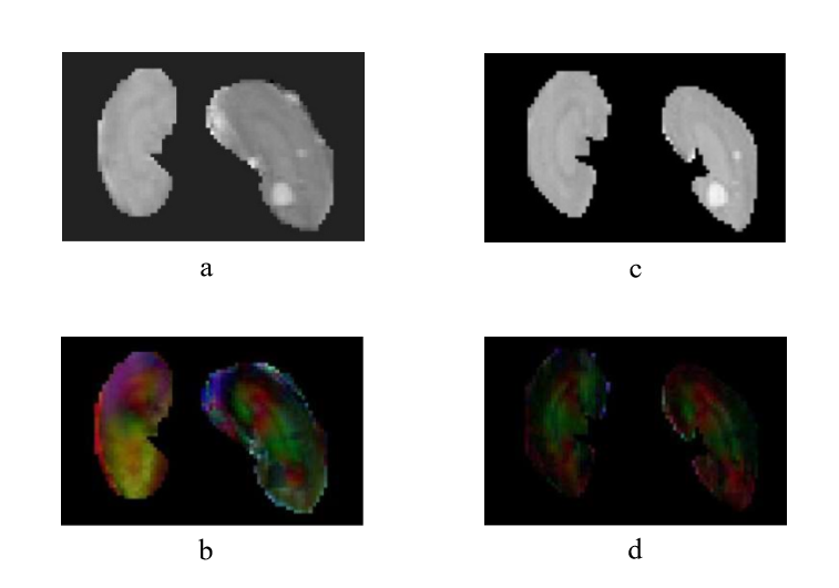
Figure
2 (a) Mean diffusivity (MD) and (b) color-encoded fractional anisotropy
(FA) map for ss-EPI. (c) MD and
(d) color-encoded FA map for 2D navigated ms-EPI.