Renat Sibgatulin1, Daniel Güllmar1, Andreas Deistung2, Stefan Ropele3, and Jürgen Rainer Reichenbach1,4,5,6
1Medical Physics Group, Institute of Diagnostic and Interventional Radiology, Jena University Hospital - Friedrich Schiller University Jena, Jena, Germany, 2Department of Radiology, University Hospital Halle (Saale), Halle, Germany, 3Department of Neurology, Medical University of Graz, Graz, Austria, 4Michael Stifel Center Jena for Data-Driven and Simulation Science, Friedrich Schiller University Jena, Jena, Germany, 5Abbe School of Photonics, Friedrich Schiller University Jena, Jena, Germany, 6Center of Medical Optics and Photonics, Friedrich Schiller University Jena, Jena, Germany
1Medical Physics Group, Institute of Diagnostic and Interventional Radiology, Jena University Hospital - Friedrich Schiller University Jena, Jena, Germany, 2Department of Radiology, University Hospital Halle (Saale), Halle, Germany, 3Department of Neurology, Medical University of Graz, Graz, Austria, 4Michael Stifel Center Jena for Data-Driven and Simulation Science, Friedrich Schiller University Jena, Jena, Germany, 5Abbe School of Photonics, Friedrich Schiller University Jena, Jena, Germany, 6Center of Medical Optics and Photonics, Friedrich Schiller University Jena, Jena, Germany
R2* anisotropy of WM is used to disentangle contributions iron and myelin in normal-appearing white matter in multiple sclerosis. Orientation independent component of R2* shows bigger difference between cohorts of MS patients and healthy controls compared to median over entire NAWM.
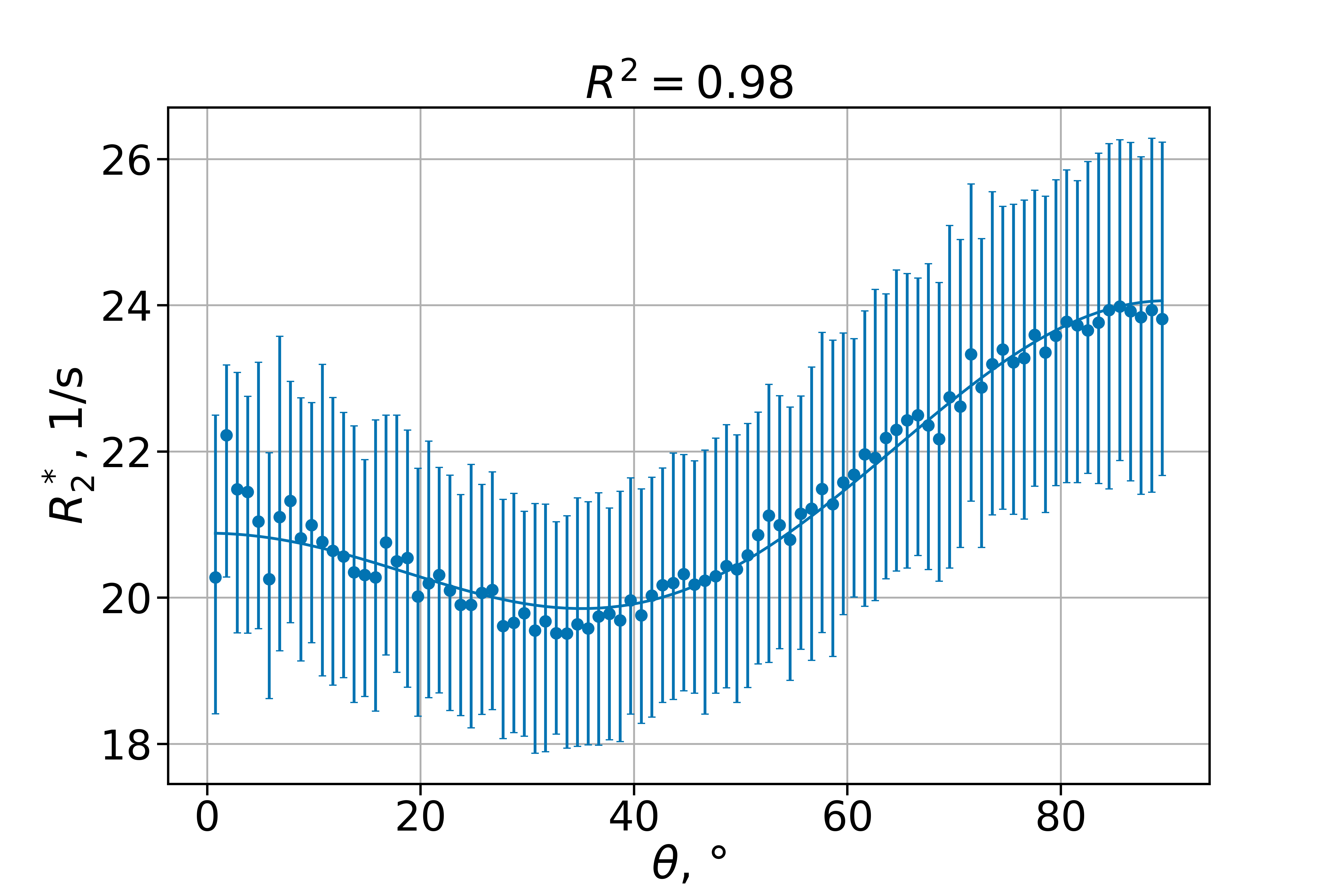
Figure 2: Orientation dependency of R2* in NAWM of an MS subject. Dots are median values within 1°-wide bins, error bars are interquartile ranges. Solid line is the fit of model 1.
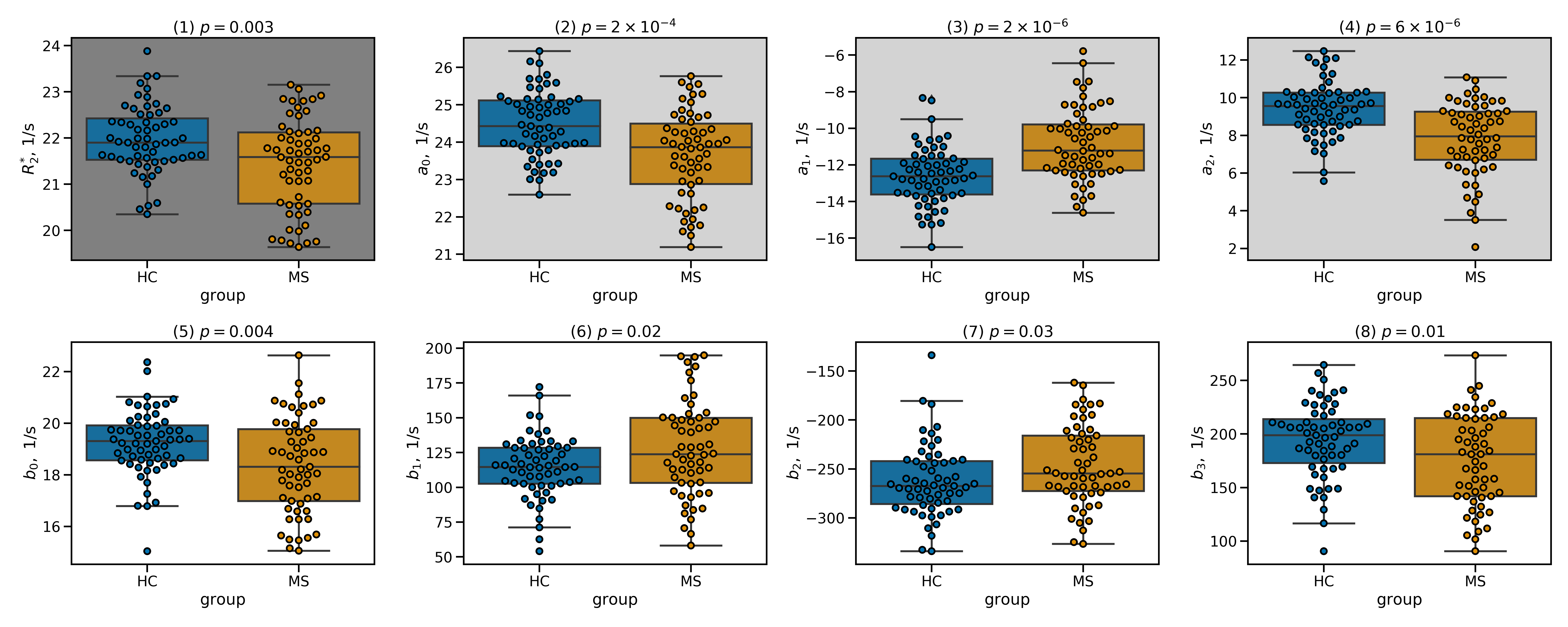
Figure 4: (1) Median R2* across the entire NAWM for healthy controls (HC) and MS patients (MS). The upper row (2–4) shows the coefficients of model 1 and the lower row (5–8) displays the coefficients of model 2 for both groups. Each dot represents one subject.