Peibei Cao1,2, Linfang Xiao1,2, Yilong Liu1,2, Yujiao Zhao1,2, Yanqiu Feng3, Alex T Leong1,2, and Ed X Wu1,2
1Laboratory of Biomedical Imaging and Signal Processing, The University of Hong Kong, Hong Kong, China, 2Department of Electrical and Electronic Engineering, The University of Hong Kong, Hong Kong, China, 3School of Biomedical Engineering, Southern Medical University, Guangzhou, China
1Laboratory of Biomedical Imaging and Signal Processing, The University of Hong Kong, Hong Kong, China, 2Department of Electrical and Electronic Engineering, The University of Hong Kong, Hong Kong, China, 3School of Biomedical Engineering, Southern Medical University, Guangzhou, China
We designed and
implemented a CNN model for partial Fourier MRI reconstruction, which
outperformed the existing projection onto convex sets (POCS) method especially
when partial Fourier fraction is close to 50%
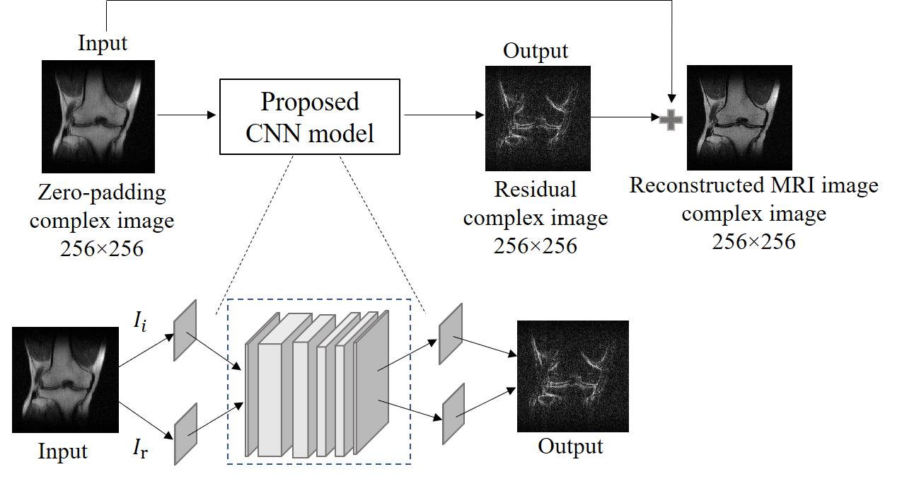
Figure 1 Illustration of the proposed CNN model architecture
and method to reconstruct MRI images. The input of the network is
a 3D matrix 256×256×2 for the image data. In this study, the k-space data has
two channels corresponding to the real and imaginary parts, respectively. The output is also a 3D matrix 256×256×2 for
the residual image. The kernel sizes of the five convolutional layers are 9×9,
7×7, 5×5, 5×5 and 3×3, respectively. The numbers of kernels are 128, 64, 32, 32
and 2, respectively. After the training, the input and output are combined for a
reconstructed MRI image.
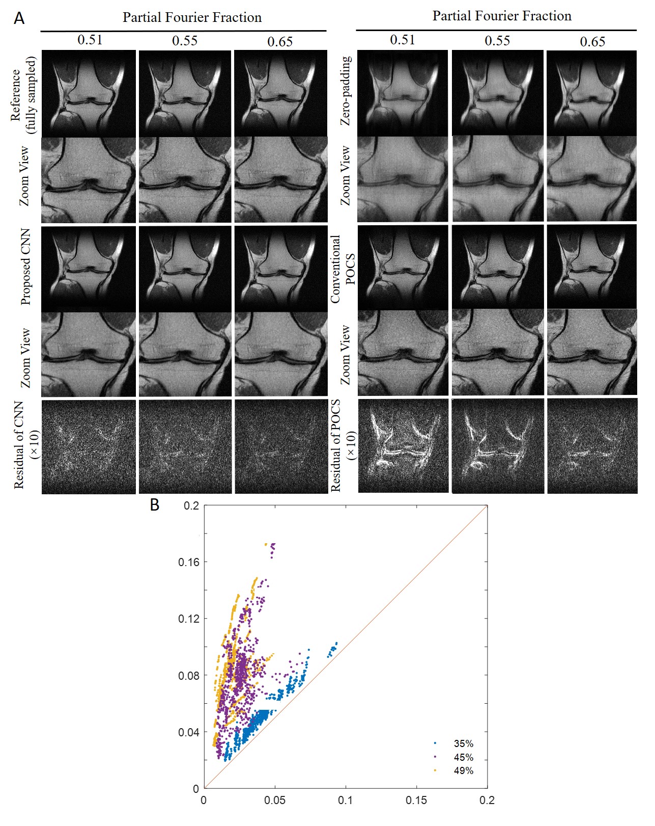
Figure 3 (A) Typical results using the CNN model, POCS, and zero-padding for different partial Fourier fractions (0.51, 0.55 and 0.65) along frequency encoding direction (vertical). POCS and CNN yielded similar performance at 0.65. However, the CNN performed better than POCS at 0.55. At 0.51, POCS suffered significant high frequency information loss, while CNN preserved sharp edges without distinct noise amplification. (B) Image residual root mean square errors (RMSEs) of the CNN and POCS calculated from 800 test images, clearly demonstrating robustness of our CNN method.