Mahdi Khajehim1,2, Thomas Christen3, Fred Tam4, Simon Graham1,4, and J. Jean Chen1,2
1Medical Biophysics, University of Toronto, Toronto, ON, Canada, 2Rotman Research Institute, Baycrest Health Sciences, Toronto, ON, Canada, 3Grenoble Institute of Neurosciences, Inserm, Grenoble, France, 4Hurvitz Brain Sciences Research Program, Sunnybrook Research Institute, Toronto, ON, Canada
1Medical Biophysics, University of Toronto, Toronto, ON, Canada, 2Rotman Research Institute, Baycrest Health Sciences, Toronto, ON, Canada, 3Grenoble Institute of Neurosciences, Inserm, Grenoble, France, 4Hurvitz Brain Sciences Research Program, Sunnybrook Research Institute, Toronto, ON, Canada
Here, a dual-stage EPI-based MRF approach with online image reconstruction is proposed for estimating T1, T2, and T2*. With the use of simultaneous multi-slice acceleration, our method can provide whole-brain coverage in less than 3 minutes.
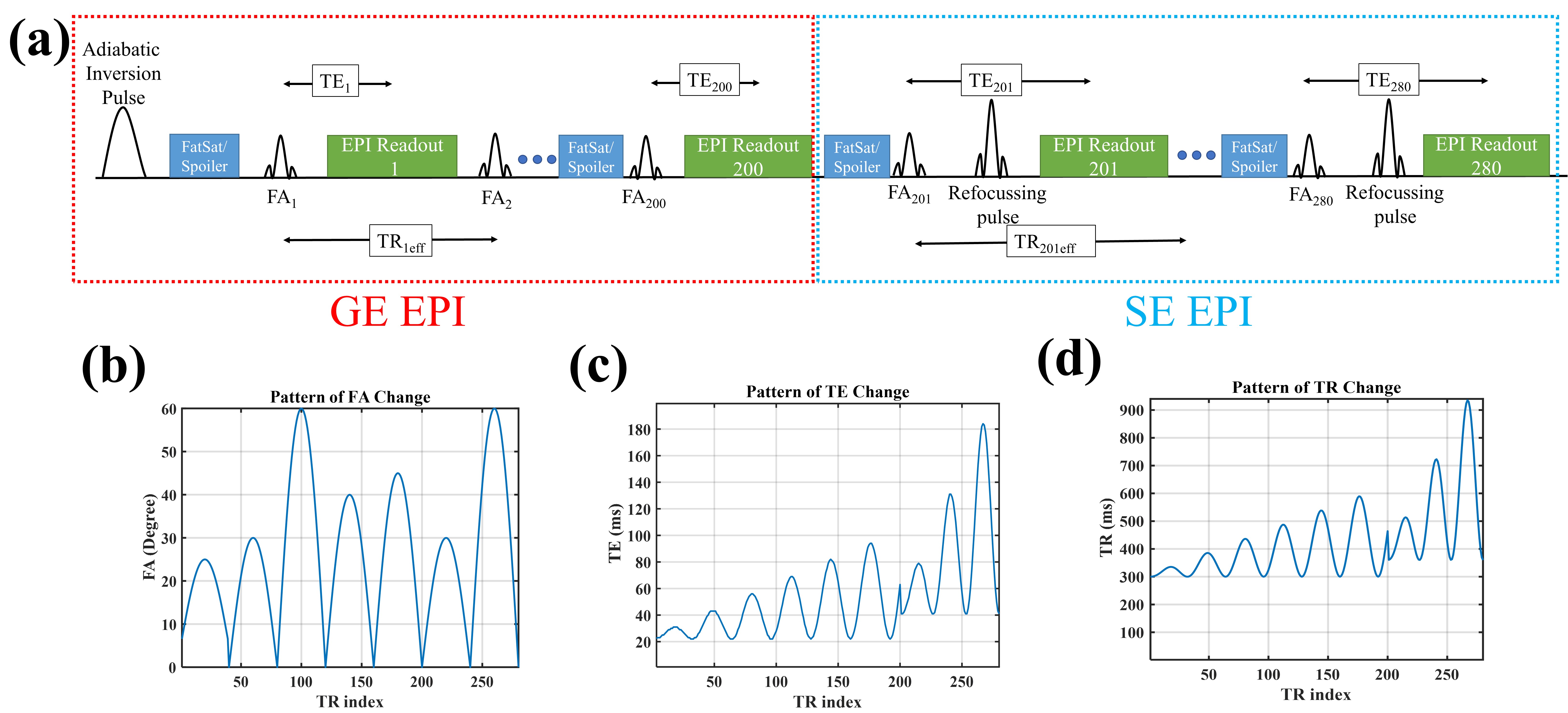
Figure 1. Schematic view of the proposed sequence, which is a combination of GE and SE EPI (a). The pattern of FA change in the sequence (b). The pattern of TE change in the sequence (c). The pattern of TR change in the sequence (d).
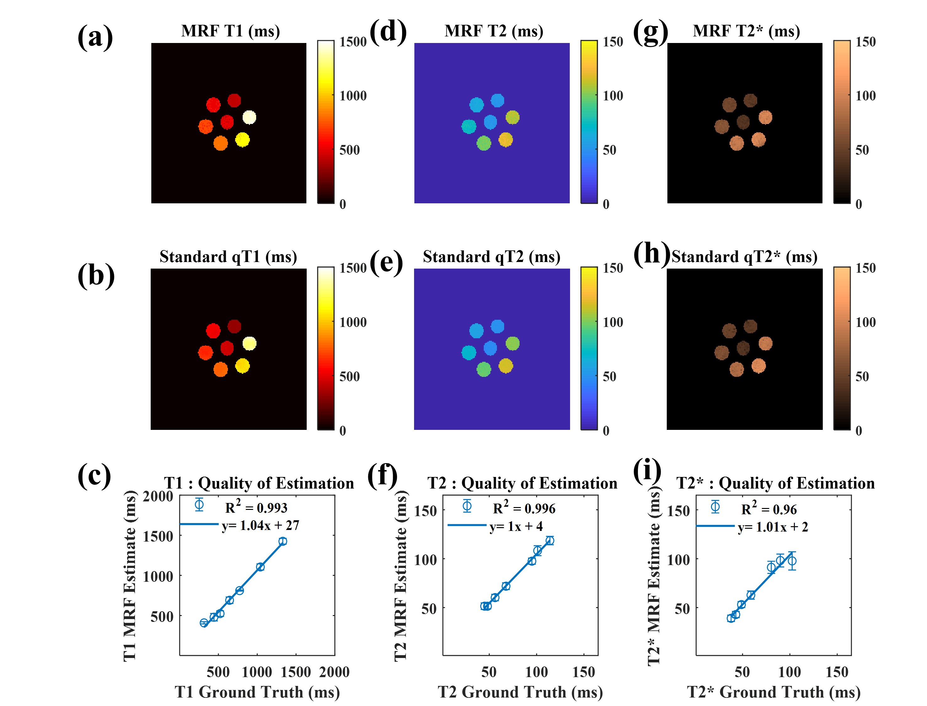
Figure 2. Phantom validation of our MRF approach based on gold-standard relaxometry methods. Strongly similar contrast was observed between our method and the validation scans (first two rows). A high degree of quantitative agreement (R2 values and regression coefficients) in the parameter estimates is shown for all cases (third row).