Yawara Haga1,2,3, Junichi Hata2,3,4, Takaaki Kaneko2,5, Tatsuhiko Yamada6, Yuji Komaki3, Fumiko Seki2,3,5, Hideyuki Okano2,5, Hirotaka James Okano4, Tetsuo Yamamori2, Noritaka Ichinohe2,7, Yuichi Yamashita7, Akira Furukawa1, and Misako Komatsu2,7
1Department of Radiological Sciences, Human Health Sciences, Tokyo Metropolitan University Graduate School, Tokyo, Japan, 2RIKEN Center for Brain Science, Saitama, Japan, 3Live Imaging Center, Central Institute for Experimental Animals, Kanagawa, Japan, 4Division of Regenerative Medicine, The Jikei University School of Medicine, Tokyo, Japan, 5Department of Physiology, Keio University School of Medicine, Tokyo, Japan, 6Graduate School of Computer Science and Systems Engineering, Kyushu Institute of Technology, Fukuoka, Japan, 7National Center of Neurology and Psychiatry, Tokyo, Japan
1Department of Radiological Sciences, Human Health Sciences, Tokyo Metropolitan University Graduate School, Tokyo, Japan, 2RIKEN Center for Brain Science, Saitama, Japan, 3Live Imaging Center, Central Institute for Experimental Animals, Kanagawa, Japan, 4Division of Regenerative Medicine, The Jikei University School of Medicine, Tokyo, Japan, 5Department of Physiology, Keio University School of Medicine, Tokyo, Japan, 6Graduate School of Computer Science and Systems Engineering, Kyushu Institute of Technology, Fukuoka, Japan, 7National Center of Neurology and Psychiatry, Tokyo, Japan
We found that some resting-state networks of awake
common marmoset brains with functional MRI and electrocorticographic methods.
Our data suggest that functional MRI data was almost consistent with electrocorticographic
data.
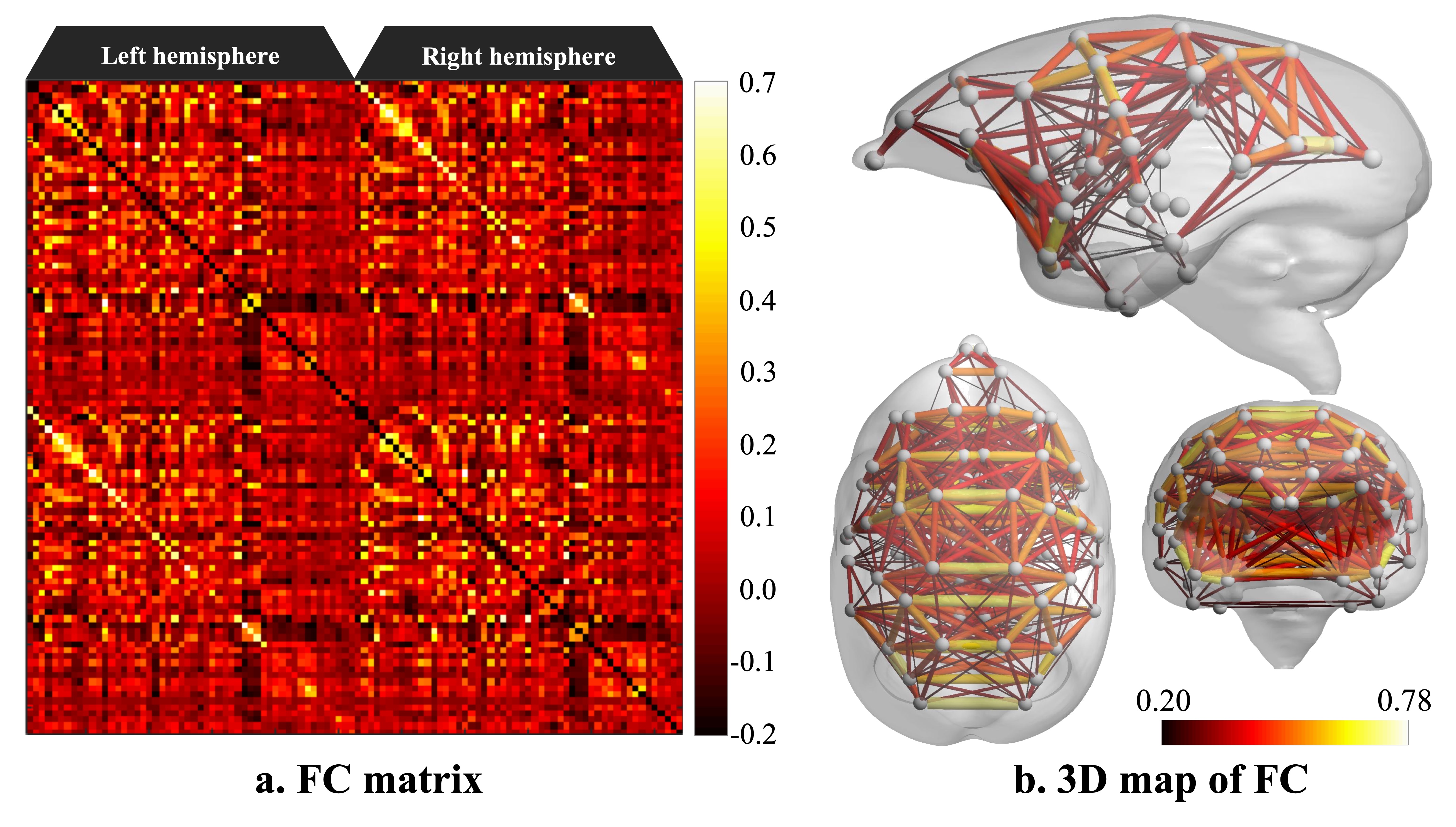
Figure 2. Functional connectivity matrix and 3D map of resting-state network in awake common marmoset
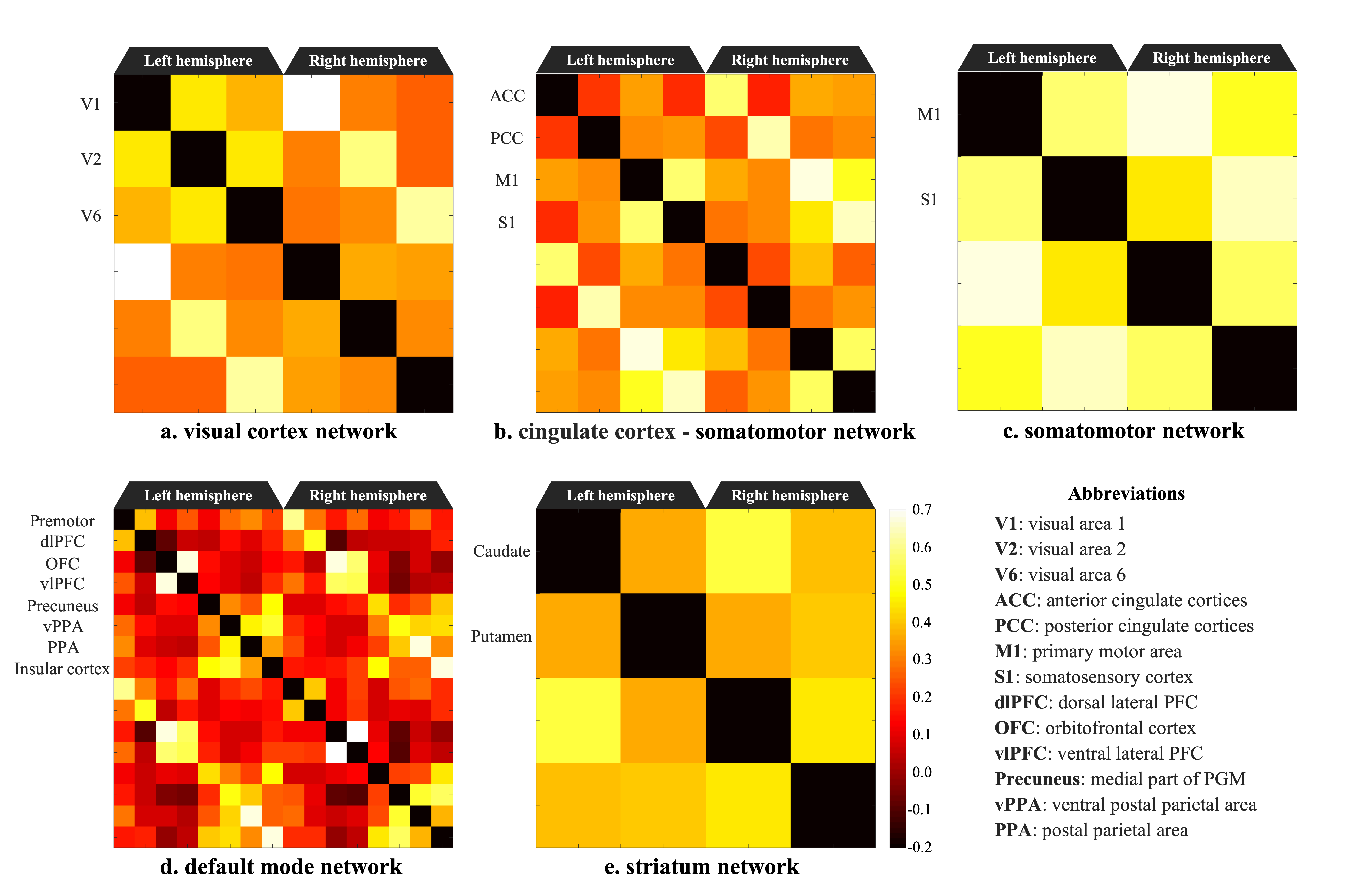
Figure 3. Resting-state network in awake common marmoset