Weiguo Li1,2, Kathleen Harris1, Ali Khan1, Simone Raiter1, Monica Matsumoto3, Andrew C Larson1, and Samdeep Mouli1
1Radiology, Northwestern University, Chicago, IL, United States, 2Research Resource Centers, University of Illinois at Chicago, Chicago, IL, United States, 3Radiology, University of Chicago, Chicago, IL, United States
1Radiology, Northwestern University, Chicago, IL, United States, 2Research Resource Centers, University of Illinois at Chicago, Chicago, IL, United States, 3Radiology, University of Chicago, Chicago, IL, United States
90Y radioembolization is a novel radiotherapy
approach that offers the potential to deliver high-dose radiation therapy with
minimal off-target toxicity. This preliminary study showed diffusion and DCE
MRI are sensitive to 90Y therapeutic effects in the
prostate.
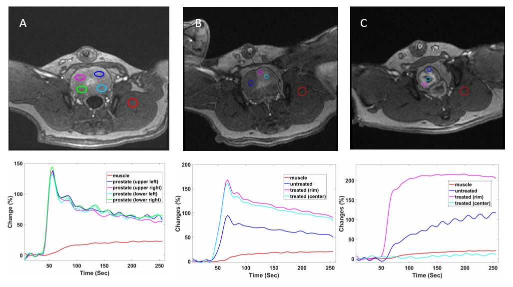
DCE
MRI images and corresponding plots of contrast enhancement versus time. A. Pre-90Y radioembolization; B. 3
days post-90Y radioembolization; C. 40 days post-90Y
radioembolization. Colored curves show percentile changes of signal intensity
versus time in the ROIs with same colors in the corresponding images.
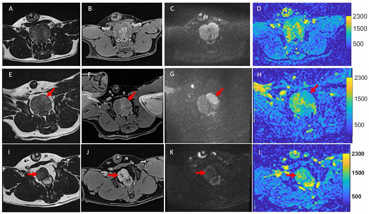
T2-weighted,
post-contrast T1-weighted, diffusion-weighted images and ADC maps for pre-90Y (top panel), 3-day post-90Y (middle
panel) and 40-day post 90Y (bottom panel). T2-weighted
images are shown in first column (A, E, I); post-contrast T1-weighted
in 2nd column (B, F J); diffusion-weighted in 3rd column
(C, G, K); and ADC maps in 4th column (D, H, L). Arrows point to the
treated lobes of the prostates.