Athanasia Kaika1,2, Mathias Schillmaier1,2, Geoffrey J. Topping1,2, and Franz Schilling1,2
1Technical University of Munich, Munich, Germany, 2Nuclear Medicine, Klinikum rechts der Isar, Munich, Germany
1Technical University of Munich, Munich, Germany, 2Nuclear Medicine, Klinikum rechts der Isar, Munich, Germany
Filter-exchange imaging (FEXI) was used to measure changes in cell
membrane permeability. AXR reduced over time upon permeabilization with ethanol,
but only minor changes in ADC, intracellular volume and Trypan staining, were
detected.
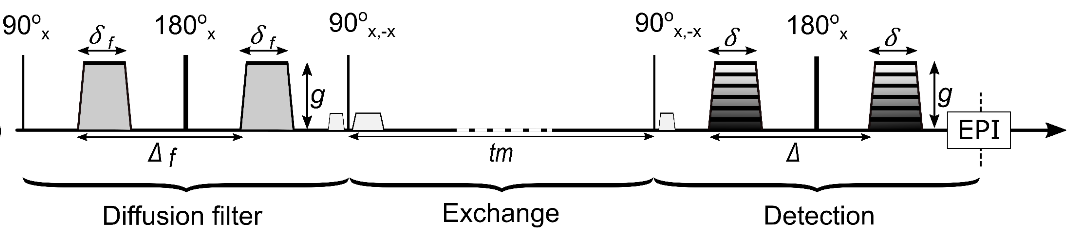
Figure 1: Schematic
representation of FEXI pulse sequence. 1. A diffusion filter module that
filters the fast diffusing component such as water in the extracellular space.
2. A storage/exchange module during which the magnetization is stored along the
longitudinal axis, during which exchange between intra and extracellular compartments
takes place. 3. A detection module including an imaging-readout. The spoiler
gradient is applied to dephase the transversal magnetization excited by the
second 90°-pulse.
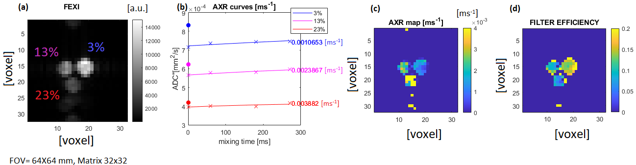
Figure 4: (a)
FEXI reference image of three yeast cell
pellets treated with 3%, 13% and 23% ethanol concentrations.
(b) AXR curves were calculated from the signal mean value in ROIs on the FEXI
images. The ROIs were drawn in the centre of each tube. (c) AXR map. (d) Filter
efficiency map.