Xinyuan Miao1,2, Yuankui Wu1,2,3, Dapeng Liu1,2, Hangyi Jiang1, Qin Qin1,2, Peter C.M van Zijl1,2, Jay J. Pillai4,5, and Jun Hua1,2
1Neurosection, Division of MRI Research, Russell H. Morgan Department of Radiology and Radiological Science, Johns Hopkins University School of Medicine, Baltimore, MD, United States, 2F.M. Kirby Research Center for Functional Brain Imaging, Kennedy Krieger Institute, Baltimore, MD, United States, 3Department of Medical Imaging, Nanfang Hospital, Southern Medical University, Guangzhou, China, 4Johns Hopkins University School of Medicine, Division of Neuroradiology, Russell H. Morgan Department of Radiology and Radiological Science, Baltimore, MD, United States, 5Department of Neurosurgery, Johns Hopkins University School of Medicine, Baltimore, MD, United States
1Neurosection, Division of MRI Research, Russell H. Morgan Department of Radiology and Radiological Science, Johns Hopkins University School of Medicine, Baltimore, MD, United States, 2F.M. Kirby Research Center for Functional Brain Imaging, Kennedy Krieger Institute, Baltimore, MD, United States, 3Department of Medical Imaging, Nanfang Hospital, Southern Medical University, Guangzhou, China, 4Johns Hopkins University School of Medicine, Division of Neuroradiology, Russell H. Morgan Department of Radiology and Radiological Science, Baltimore, MD, United States, 5Department of Neurosurgery, Johns Hopkins University School of Medicine, Baltimore, MD, United States
Diffusion-prepared
diffusion tensor imaging with three-dimensional fast gradient-echo readout can significantly
reduce susceptibility artifacts commonly seen in conventional spin-echo echo-planar-imaging
diffusion tensor imaging in the presence of metallic orthodontic braces.
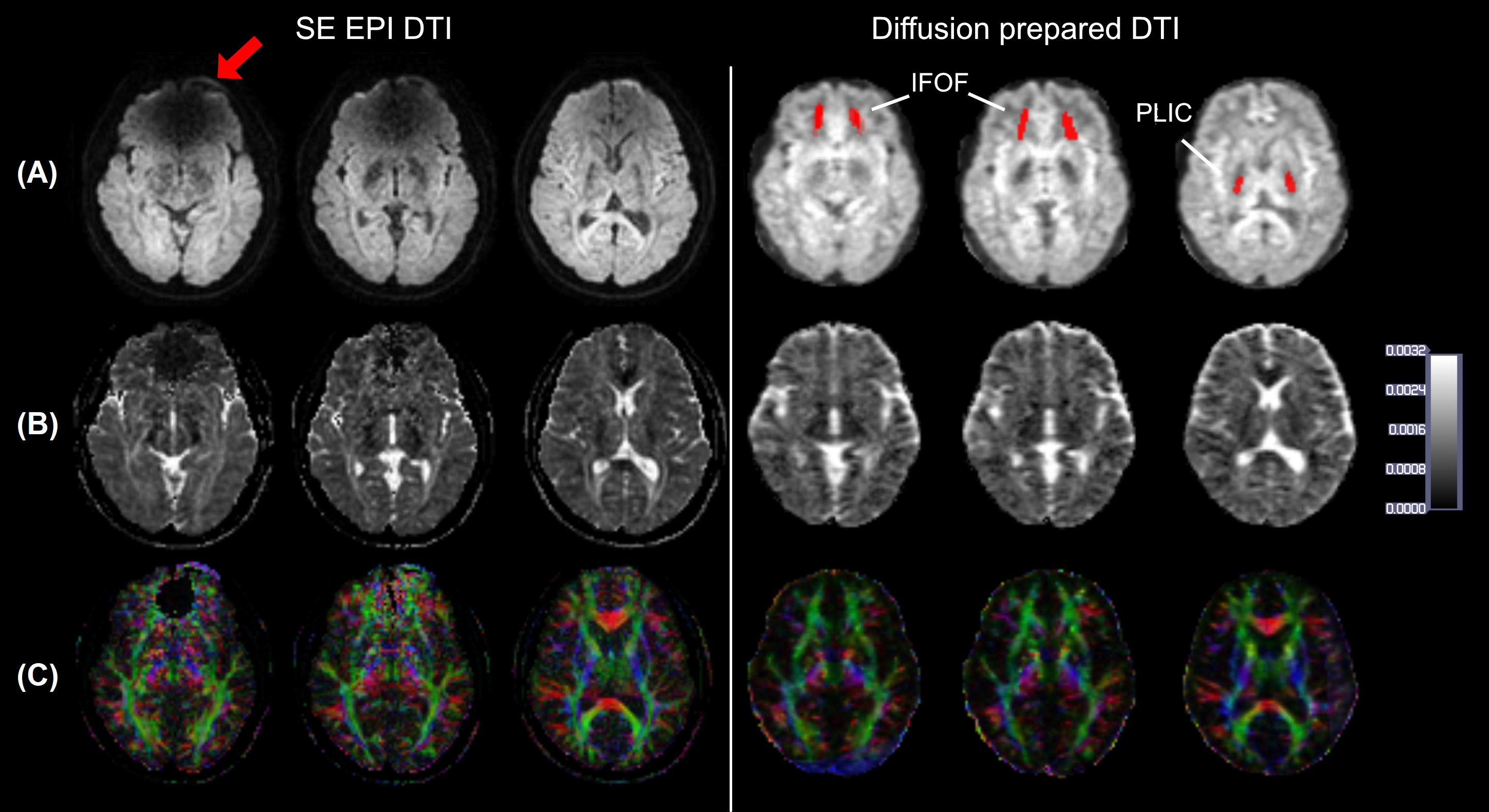
Figure 2. SE-EPI and diffusion-prepared DTI axial images acquired at 3.0 T in a participant wearing metallic dental
braces. A, raw diffusion-weighted images, B, calculated ADC maps, and, C, fractional anisotropy map color
coded by V1 orientation (standard red, green, and blue convention).
Susceptibility artifacts were observed on SE EPI images in regions close to
brace (arrow). No obvious artifacts were seen on diffusion-prepared DTI image.
Regions of interest of IFOF and PLIC used in subsequent quantitative
analysis are highlighted on diffusion-prepared DTI images with red.

Figure 3. Geometric distortions in axial SE-EPI (top row) and
diffusion-prepared (bottom row) DTI when compared with MPRAGE images
in same participant wearing metallic dental braces. Edges of brain structures
obtained from coregistered MPRAGE images are shown in red contour lines on mean
diffusion-weighted images from the two DTI approaches. Mismatch between
contour lines and edge of structures shown in DTI images illustrates geometric
distortion artifacts (eg, frontal area indicated by arrow). Mean Jaccard index
(JI) is calculated for each slice and is listed under each image.