Lebina Shrestha Kakkar1, Muhammad Usman1, Alex Kirkham2, Simon Arridge1, and David Atkinson1
1Centre for Medical Imaging, University College London, London, United Kingdom, 2Department of Radiology, University College Hospital, London, United Kingdom
1Centre for Medical Imaging, University College London, London, United Kingdom, 2Department of Radiology, University College Hospital, London, United Kingdom
Distortion correction of a clinical standard
diffusion-weighted prostate MRI is feasible using model-based reconstruction.
An increase in SNR of averaged high b-value data is a complimentary product of
this framework.

T2W image (1st column), ADC maps computed from corrected (2nd column) and uncorrected (3rd column) DW datasets are displayed here for two patients. Dice score of the prostate ROI between the T2W image and the corrected ADC, and the T2W image and the uncorrected standard ADC are: 0.95 and 0.86 for patient 1, and 0.89 and 0.87 for patient 2. The current framework performs slightly better than the uncorrected DW data both in terms of Dice scores and discriminating between prostatic zones with respect to the T2W image.
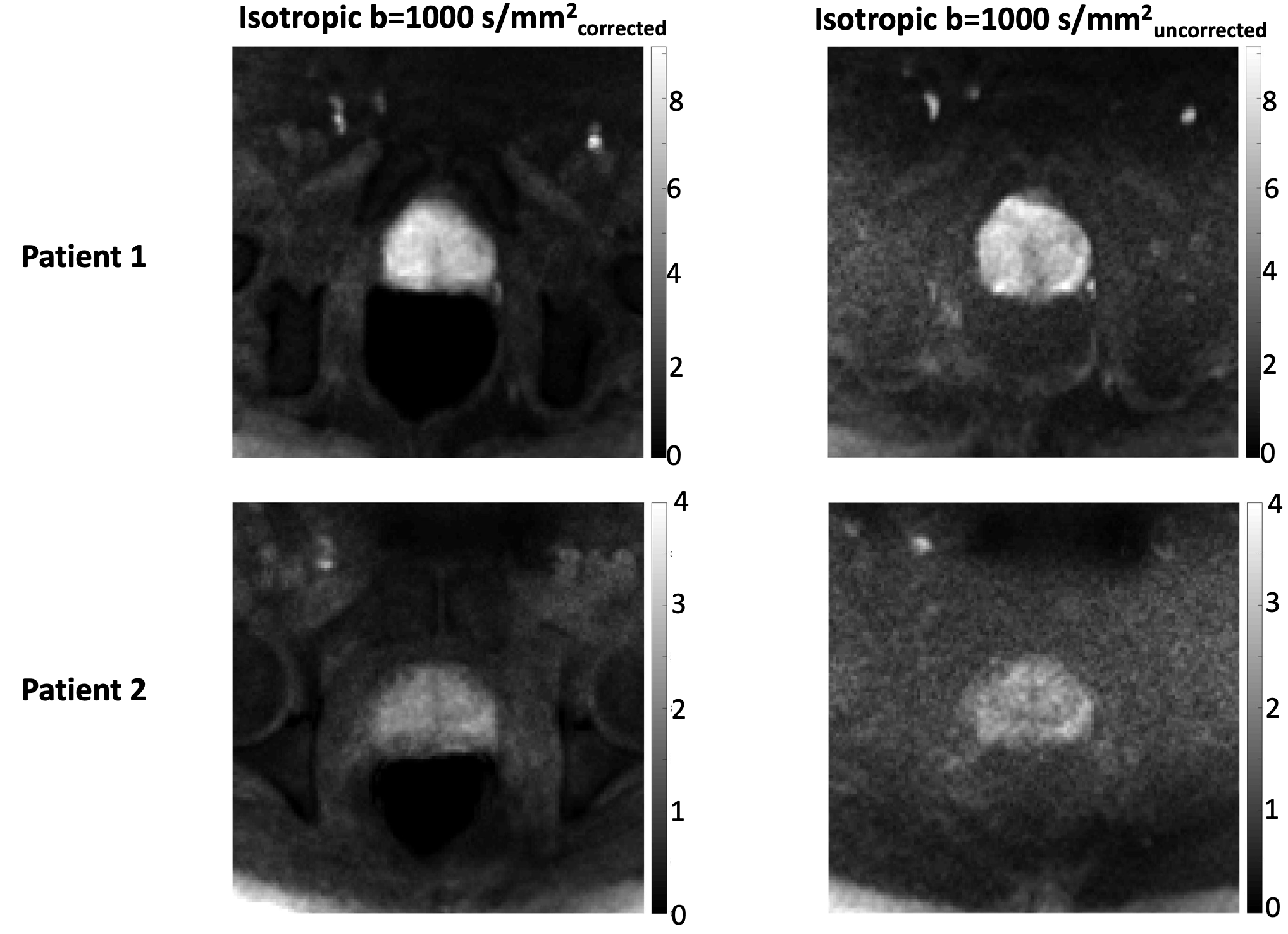
Corrected (1st column) and uncorrected (2nd column) isotropically averaged b=1000 s/mm2
for the same slices as in Fig.2 of the same two patients are shown here. The
mean and standard deviation of image SNR for the displayed slice for Patient 1
are: 7.2±1.3 (corrected data) and 3.9±0.5 (uncorrected data). For patient 2 the
image SNR values are: 3.0±0.3 (corrected data) and 2.1±0.4 (uncorrected data).