Vincent Kyu Lee1, Benjamin Meyers1, William T Reynolds2, Rafael Ceschin1, Vincent Schmithorst1, Jeffrey Berman3, Thomas Chenevert4, Borjan Gagoski5, Peter LaViolette6, Deqiang Qiu7, Sudhir Pathak1, Ashok Panigrahy1, and Walter Schneider8
1Radiology, University of Pittsburgh, Pittsburgh, PA, United States, 2Biomedical Informatics, University of Pittsburgh, Pittsburgh, PA, United States, 3Radiology, Children's Hospital of Philadelphia, Philadelphia, PA, United States, 4Radiology, University of Michigan, Ann Arbor, MI, United States, 5Radiology, Harvard Medical School, Boston, MA, United States, 6Radiology, Medical College of Wisconsin, Milwaukee, WI, United States, 7Radiology, Emory University, Atlanta, GA, United States, 8Psychology, University of Pittsburgh, Pittsburgh, PA, United States
1Radiology, University of Pittsburgh, Pittsburgh, PA, United States, 2Biomedical Informatics, University of Pittsburgh, Pittsburgh, PA, United States, 3Radiology, Children's Hospital of Philadelphia, Philadelphia, PA, United States, 4Radiology, University of Michigan, Ann Arbor, MI, United States, 5Radiology, Harvard Medical School, Boston, MA, United States, 6Radiology, Medical College of Wisconsin, Milwaukee, WI, United States, 7Radiology, Emory University, Atlanta, GA, United States, 8Psychology, University of Pittsburgh, Pittsburgh, PA, United States
Our study showed synthetic phantoms that simulate fiber anatomical characteristics can reduce cross
scanner variability and can provide in vitro corrections factors for reducing in-vivo inter-scanner variance specific to discrete segments of cortical association fiber tracts.
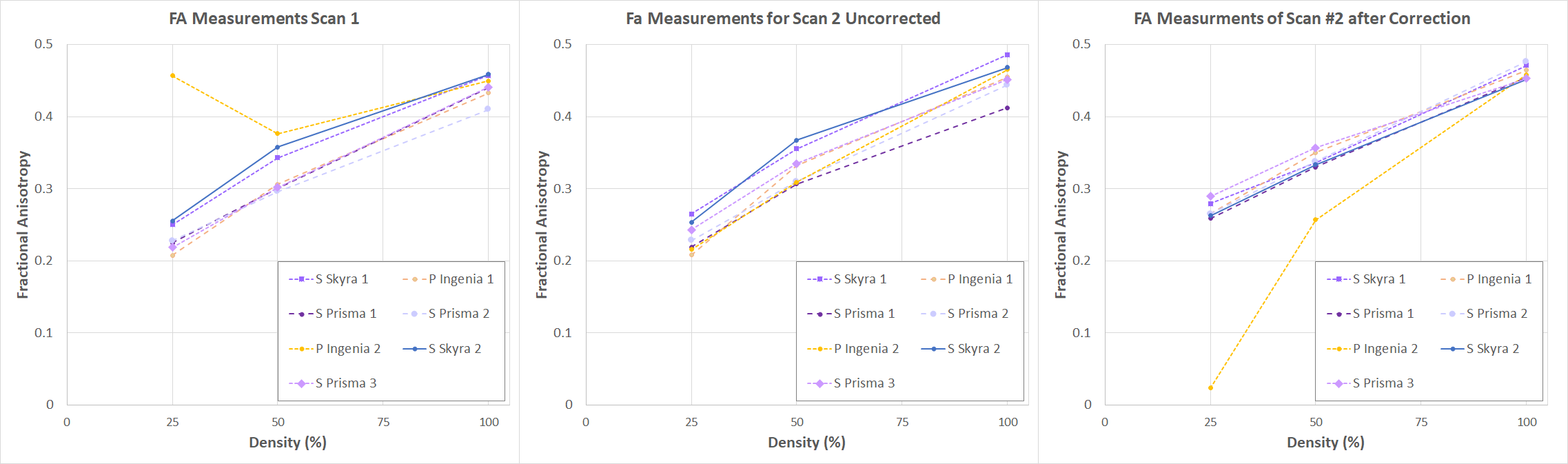
Figure 2. Synthetic phantom correction calculations using FA measurements from DTI imaging of the synthetic phantom unidirectional fiber blocks. The first set of scans (A) were used to determine the scanner specific correction factors which were then applied to the second set of scans.For all scanners (B). After the correction (C), a reduction in cross scanner variability is observed.
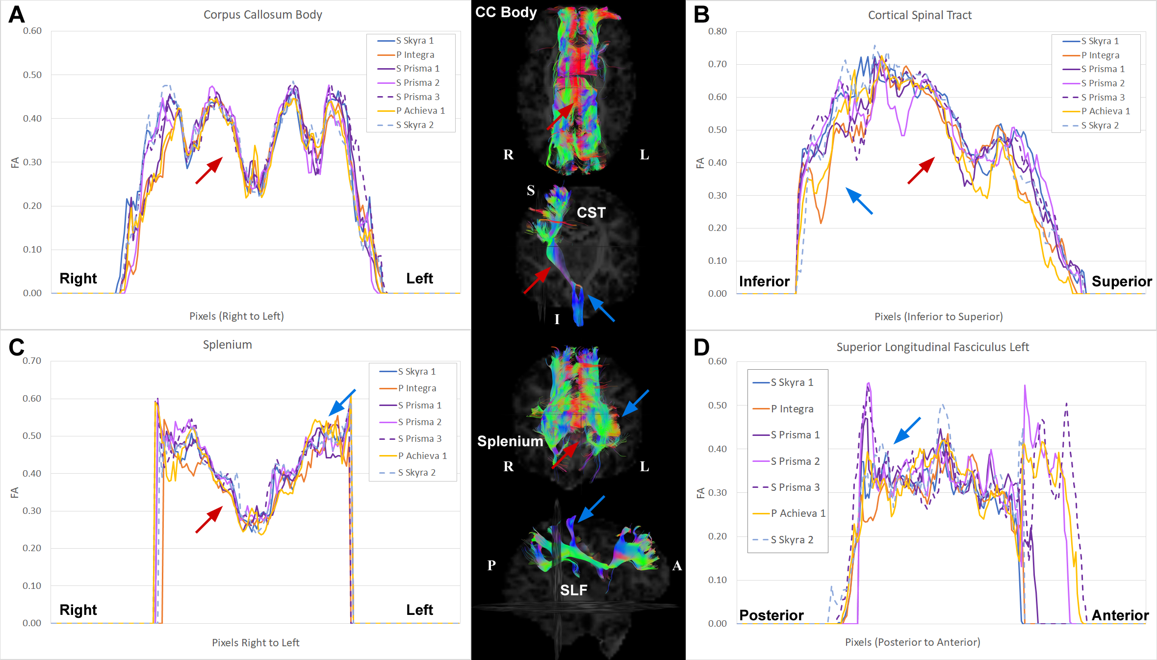
Figure 3. Along-tract FA analysis
of an individual human phantom across multiple sites. The tracts presented are
CC Body (A), right CST (B), Splenium (C), and left SLF (D). Present to varying degrees in all the tracts
are regions that are more sensitive to scanner variability (blue arrows) and
regions that are more reliably measured (red arrows) regardless of MRI used.