Junqi Xu1, He Wang1,2, Xueying Zhao1, Hui Zhang1, Xiaoyuan Feng3, and Ren Yan3
1Institute of Science and Technology for Brain-Inspired Intelligence, Fudan University, Shanghai, China, 2Human Phenome Institute, Fudan University, Shanghai, China, 3Radiology, Huashan Hospital, Fudan University, Shanghai, China
1Institute of Science and Technology for Brain-Inspired Intelligence, Fudan University, Shanghai, China, 2Human Phenome Institute, Fudan University, Shanghai, China, 3Radiology, Huashan Hospital, Fudan University, Shanghai, China
We
have developed a multi-model of DWI platform for pathological diagnosis. Herein,
FROC, CTRW and DKI performed best among these models in differentiating low-
and high-grade adults brain tumor.
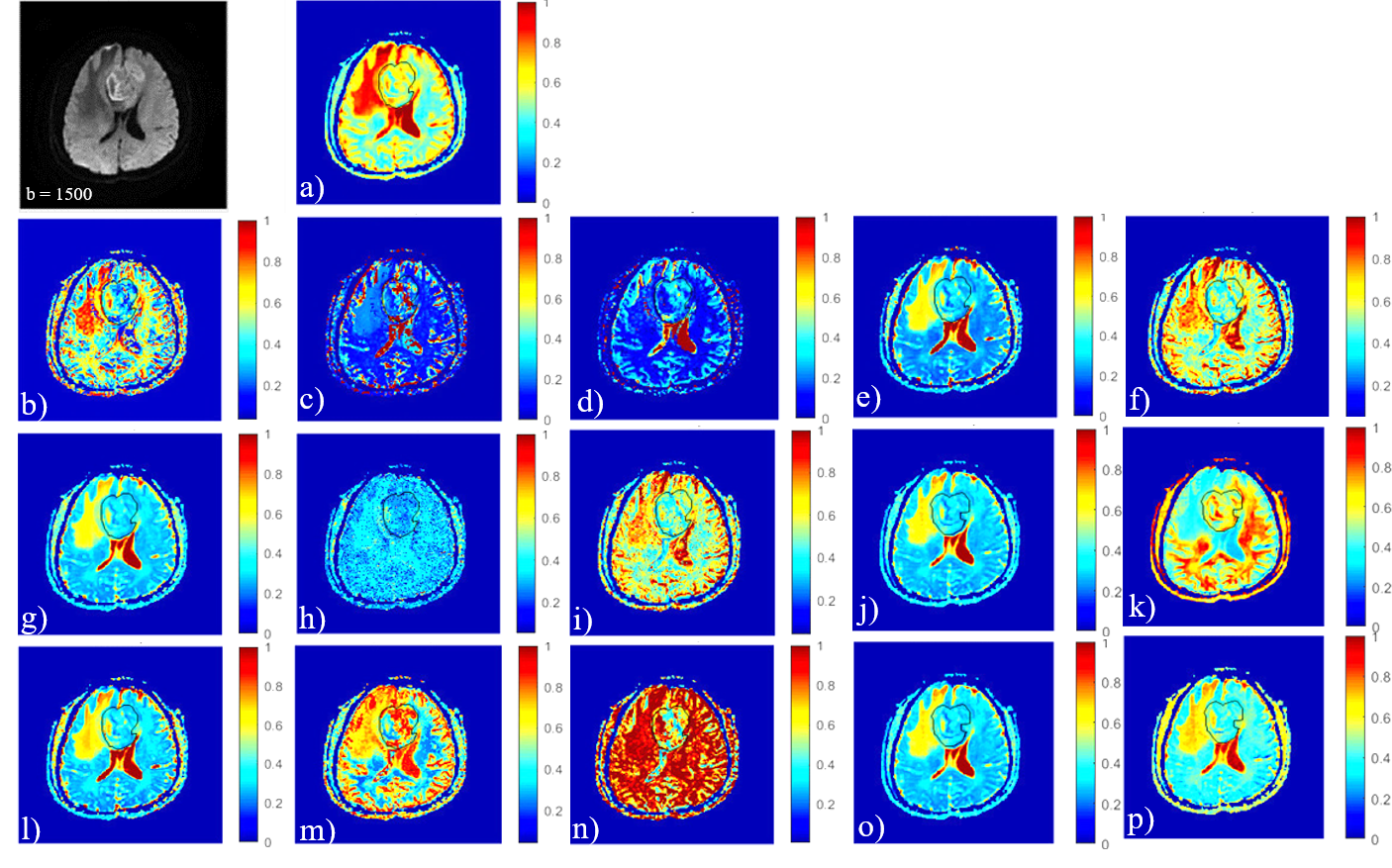
Fig. 1 All models of diffusion MR imaging. This image
shows one patient with high-grade brain tumor, where the first image is DWI at b = 1500s/mm2and from image
a) to p) represents "ADCmap", "Dmap", "Df map", "Dsmap" of IVIM ,"DDCmap","$$$\alpha$$$map" of SEM,"Dmap","$$$\mu$$$map","$$$\beta_f$$$map" of FROC,"Dcmap","$$$\alpha_c$$$map","$$$\beta_c$$$map" of CTRW, "Dk map","Kmap"of DKI and "ADCSmap ” of SM respectively.
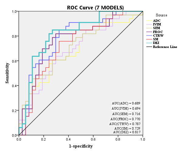
Fig.
2
Jointly parameters in each models and this picture shows ROC analysis of 7
models.