Timo Roine1,2, Oskari Kantonen3, Ulrika Roine1, Sami Virtanen4, Jani Saunavaara4,5, Riitta Parkkola4, Ruut Laitio6, Olli Arola6, Marja Hynninen7, Juha Martola8, Heli M Silvennoinen8, Marjaana Tiainen9, Risto O. Roine10, Harry Scheinin6, and Timo Laitio6
1Department of Neuroscience and Biomedical Engineering, Aalto University School of Science, Espoo, Finland, 2Turku Brain and Mind Center, University of Turku, Turku, Finland, 3Turku PET Centre, University of Turku and the Hospital District of Southwest Finland, Turku, Finland, 4Department of Radiology, Turku University Hospital, University of Turku, Turku, Finland, 5Department of Medical Physics, Turku University Hospital, University of Turku, Turku, Finland, 6Division of Perioperative Services, Intensive Care and Pain Medicine, Turku University Hospital, University of Turku, Turku, Finland, 7Division of Intensive Care Medicine, University of Helsinki and Helsinki University Hospital, Helsinki, Finland, 8Department of Radiology, University of Helsinki and Helsinki University Hospital, Helsinki, Finland, 9Department of Neurology, University of Helsinki and Helsinki University Hospital, Helsinki, Finland, 10Division of Clinical Neurosciences, Turku University Hospital, University of Turku, Turku, Finland
1Department of Neuroscience and Biomedical Engineering, Aalto University School of Science, Espoo, Finland, 2Turku Brain and Mind Center, University of Turku, Turku, Finland, 3Turku PET Centre, University of Turku and the Hospital District of Southwest Finland, Turku, Finland, 4Department of Radiology, Turku University Hospital, University of Turku, Turku, Finland, 5Department of Medical Physics, Turku University Hospital, University of Turku, Turku, Finland, 6Division of Perioperative Services, Intensive Care and Pain Medicine, Turku University Hospital, University of Turku, Turku, Finland, 7Division of Intensive Care Medicine, University of Helsinki and Helsinki University Hospital, Helsinki, Finland, 8Department of Radiology, University of Helsinki and Helsinki University Hospital, Helsinki, Finland, 9Department of Neurology, University of Helsinki and Helsinki University Hospital, Helsinki, Finland, 10Division of Clinical Neurosciences, Turku University Hospital, University of Turku, Turku, Finland
We investigated structural brain connectivity networks after
out-of-hospital cardiac arrest to detect differences related to survival. We
found that decreased strength and efficiency were both globally and locally related
to increased mortality at 6 months.
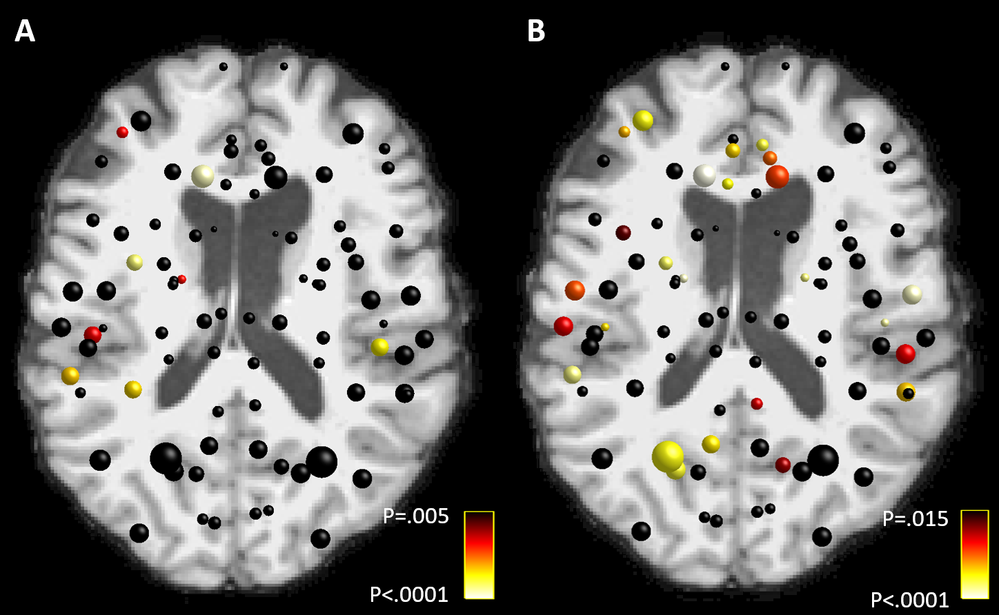
Figure
2. Local decreases in A) strength and B) local
efficiency of the structural brain connectivity networks in patients who did
not survive after OHCA compared to the survivors. The color of the node
indicates the statistical significance of the results. The results were adjusted for false discovery rate (FDR) with a
significance threshold 0.05. Age, gender, and imaging site were used as
covariates in the statistical model.
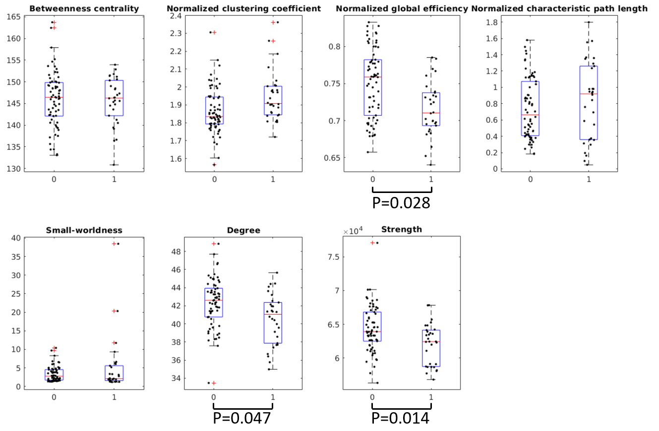
Figure 1. Global structural
brain connectivity network differences between the survivors (group 0) and
those who did not survive (group 1) after OHCA. The P-values were adjusted for
false discovery rate (FDR). Age, gender, and imaging site were used as
covariates in the statistical model.