Steven Jillings1, Angelique Van Ombergen2, Elena Tomilovskaya3, Alena Rumshiskaya4, Liudmila Litvinova4, Inna Nosikova3, Ekaterina Pechenkova5, Ilya Rukavishnikov3, Inessa Kozlovskaya3, Stefan Sunaert6, Paul M Parizel7, Valentin Sinitsyn8, Victor Petrovichev4, Steven Laureys9, Peter zu Eulenburg10, Jan Sijbers11, Floris Wuyts1, and Ben Jeurissen11
1Lab for Equilibrium Investigations and Aerospace, Dept. of Physics, University of Antwerp, Antwerp, Belgium, 2Translations Neuroscience, Dept. of Medicine, University of Antwerp, Antwerp, Belgium, 3Institute of Biomedical Problems, Russian Academy of Sciences, Moscow, Russian Federation, 4Dept. of Radiology, Federal Center of Treatment and Rehabilitation, Moscow, Russian Federation, 5Laboratory for Cognitive Research, National Research University Higher School of Economics, Moscow, Russian Federation, 6Dept. of Imaging and Pathology, KU Leuven, Leuven, Belgium, 7Dept. of Radiology, Royal Perth Hospital and University of Western Australia, Perth, Australia, 8Faculty of Fundamental Medicine, Lomonosov Moscow State University, Moscow, Russian Federation, 9Coma Science Group, Dept. of Neurology, University (Hospital) of Liège, Liège, Belgium, 10German Center for Vertigo and Balance Disorders, Dept. of Neurology, Ludwig-Maximilians-University Munich, Munich, Germany, 11imec - Vision Lab, Dept. of Physics, University of Antwerp, Antwerp, Belgium
1Lab for Equilibrium Investigations and Aerospace, Dept. of Physics, University of Antwerp, Antwerp, Belgium, 2Translations Neuroscience, Dept. of Medicine, University of Antwerp, Antwerp, Belgium, 3Institute of Biomedical Problems, Russian Academy of Sciences, Moscow, Russian Federation, 4Dept. of Radiology, Federal Center of Treatment and Rehabilitation, Moscow, Russian Federation, 5Laboratory for Cognitive Research, National Research University Higher School of Economics, Moscow, Russian Federation, 6Dept. of Imaging and Pathology, KU Leuven, Leuven, Belgium, 7Dept. of Radiology, Royal Perth Hospital and University of Western Australia, Perth, Australia, 8Faculty of Fundamental Medicine, Lomonosov Moscow State University, Moscow, Russian Federation, 9Coma Science Group, Dept. of Neurology, University (Hospital) of Liège, Liège, Belgium, 10German Center for Vertigo and Balance Disorders, Dept. of Neurology, Ludwig-Maximilians-University Munich, Munich, Germany, 11imec - Vision Lab, Dept. of Physics, University of Antwerp, Antwerp, Belgium
Spaceflight causes a redistribution of CSF and concomitant GM density changes. As opposed to earlier findings, we can strongly conclude that the GM changes are morphological and do not point to atrophy. The net amount of GM and WM increased in sensorimotor areas, indicating neuroplasticity.
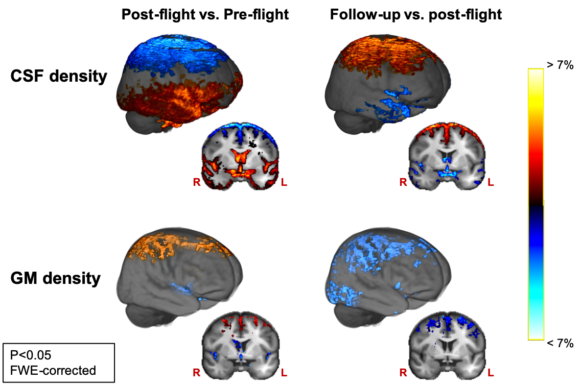
Fig 2: CSF and GM density change in the opposite direction at the superior frontal and parietal part of the cerebrum, and along the ventricles and the Sylvian fissure between pre-flight and post-flight. These changes are then largely reversed seven months after spaceflight as revealed by the comparison of follow-up to post-flight. Results are scaled by effect size and are overlayed onto the group template image.
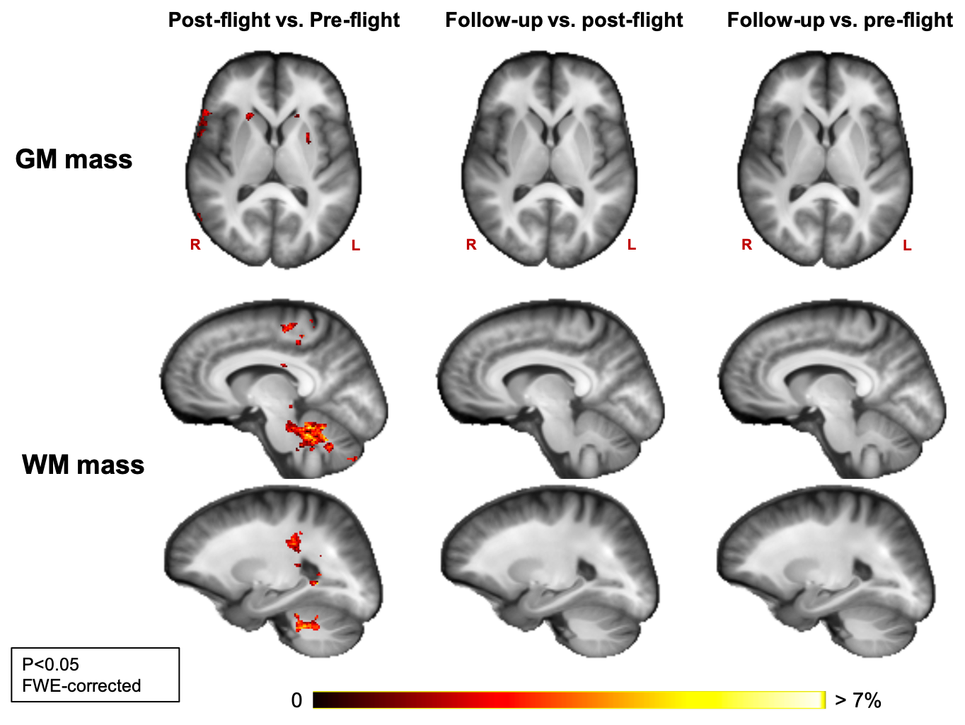
Fig 1: GM mass increases from pre- to post-flight are found in the basal ganglia. WM mass increased from pre- to post-flight in the cerebellum, in a part of the corticospinal tract and in the pre- and postcentral gyri. No significant decreases in GM or WM tissue mass were found between pre- and post-flight. No significant changes were observed between post-flight and follow-up, and between pre-flight and follow-up. Results are scaled by effect size and are overlayed onto the group template image.