Graham A. D. Archibald1, Jordan A. Chad1,2, David H. Salat3,4, and J. Jean Chen1,2
1Rotman Research Institute, Baycrest Health Sciences, Toronto, ON, Canada, 2Department of Medical Biophysics, University of Toronto, Toronto, ON, Canada, 3MGH/HST Athinoula A. Martinos Center for Biomedical Imaging, Massachusetts General Hospital, Harvard Medical School, Charlestown, MA, United States, 4Neuroimaging Research for Veterans Center, VA Boston Healthcare System, Boston, MA, United States
1Rotman Research Institute, Baycrest Health Sciences, Toronto, ON, Canada, 2Department of Medical Biophysics, University of Toronto, Toronto, ON, Canada, 3MGH/HST Athinoula A. Martinos Center for Biomedical Imaging, Massachusetts General Hospital, Harvard Medical School, Charlestown, MA, United States, 4Neuroimaging Research for Veterans Center, VA Boston Healthcare System, Boston, MA, United States
Mean diffusivity (MD), derived from diffusion MRI, serves as a more sensitive measure of aging than thickness across the cortex. MD shows distinct effects than thickness, suggesting that MD provides insight into microscopic degeneration that cannot be detected with macroscopic MRI measures.
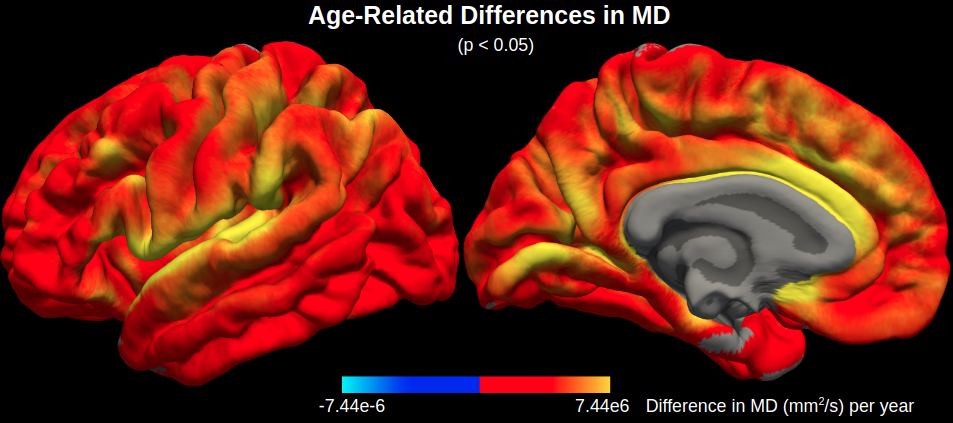
Figure 1. Age-related differences in mean diffusivity (MD). MD is significantly correlated with age across the cerebral cortex, particularly in the cingulate, insular and superior temporal cortices.
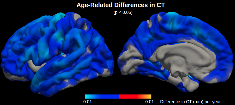
Figure 2. Age-related differences in cortical thickness (CT). CT is significantly correlated with age across the cerebral cortex, particularly in the motor cortex, although there are less significant regions with age-related differences in CT than there are in MD.