Madeleine T Dacey1,2,3, Stefan E Poirier1,3, Janice Gomes2,4, Udunna C Anazodo1,3, and Christopher W McIntyre1,2
1Medical Biophysics, Western University, London, ON, Canada, 2Kidney Clinical Research Unit, Lawson Health Sciences Center, London, ON, Canada, 3Imaging, Lawson Health Research Insitute, London, ON, Canada, 4Pathology and Laboratory Medicine, Western University, London, ON, Canada
1Medical Biophysics, Western University, London, ON, Canada, 2Kidney Clinical Research Unit, Lawson Health Sciences Center, London, ON, Canada, 3Imaging, Lawson Health Research Insitute, London, ON, Canada, 4Pathology and Laboratory Medicine, Western University, London, ON, Canada
Cognitive impairment and white matter degeneration are common in hemodialysis (HD) patients. To identify the acute effects of HD on the brain, we used a novel system to perform MRI scans during HD. The results indicate ischemia and osmotic imbalances cause acute brain injury.

White and grey matter volume increase significantly at peak stress during hemodialysis while cerebrospinal fluid volume decreases (p<0.05). These changes are consistent with brain swelling and indicate ionic edema.
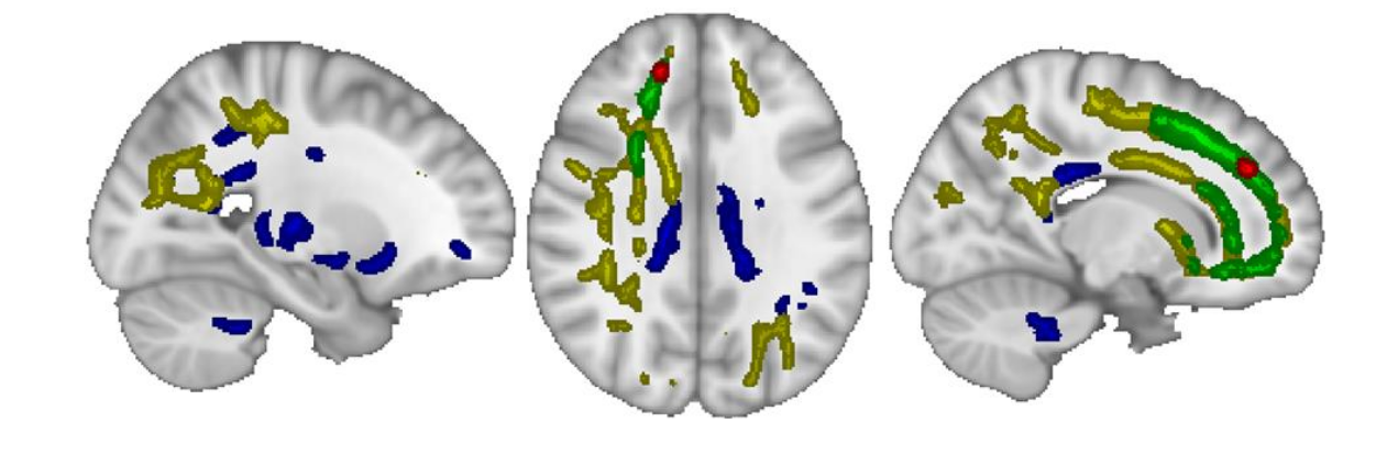
The colored regions show where each diffusion metric increased significantly (P<0.05) at peak stress during hemodialysis. The blue and yellow regions correspond to fractional anisotropy and axial diffusivity, respectively, and indicate cytotoxic edema. The green and red regions correspond to mean diffusivity. Radial diffusivity is shown in red.