Shana Black1,2, Andrew Janson1,2, and Christopher R Butson1,2,3
1Biomedical Engineering, University of Utah, Salt Lake City, UT, United States, 2Scientific Computing and Imaging Institute, University of Utah, Salt Lake City, UT, United States, 3Neurology, Neurosurgery, and Psychiatry, University of Utah, Salt Lake City, UT, United States
1Biomedical Engineering, University of Utah, Salt Lake City, UT, United States, 2Scientific Computing and Imaging Institute, University of Utah, Salt Lake City, UT, United States, 3Neurology, Neurosurgery, and Psychiatry, University of Utah, Salt Lake City, UT, United States
Statistically significant (FWE-corrected p<0.1)
increases in FC and FDC are evident in a commissural pathway including the
splenium and regions of the retrosplenial complex in SCI subjects with chronic
neuropathic pain when compared to SCI subjects without any pain symptoms.
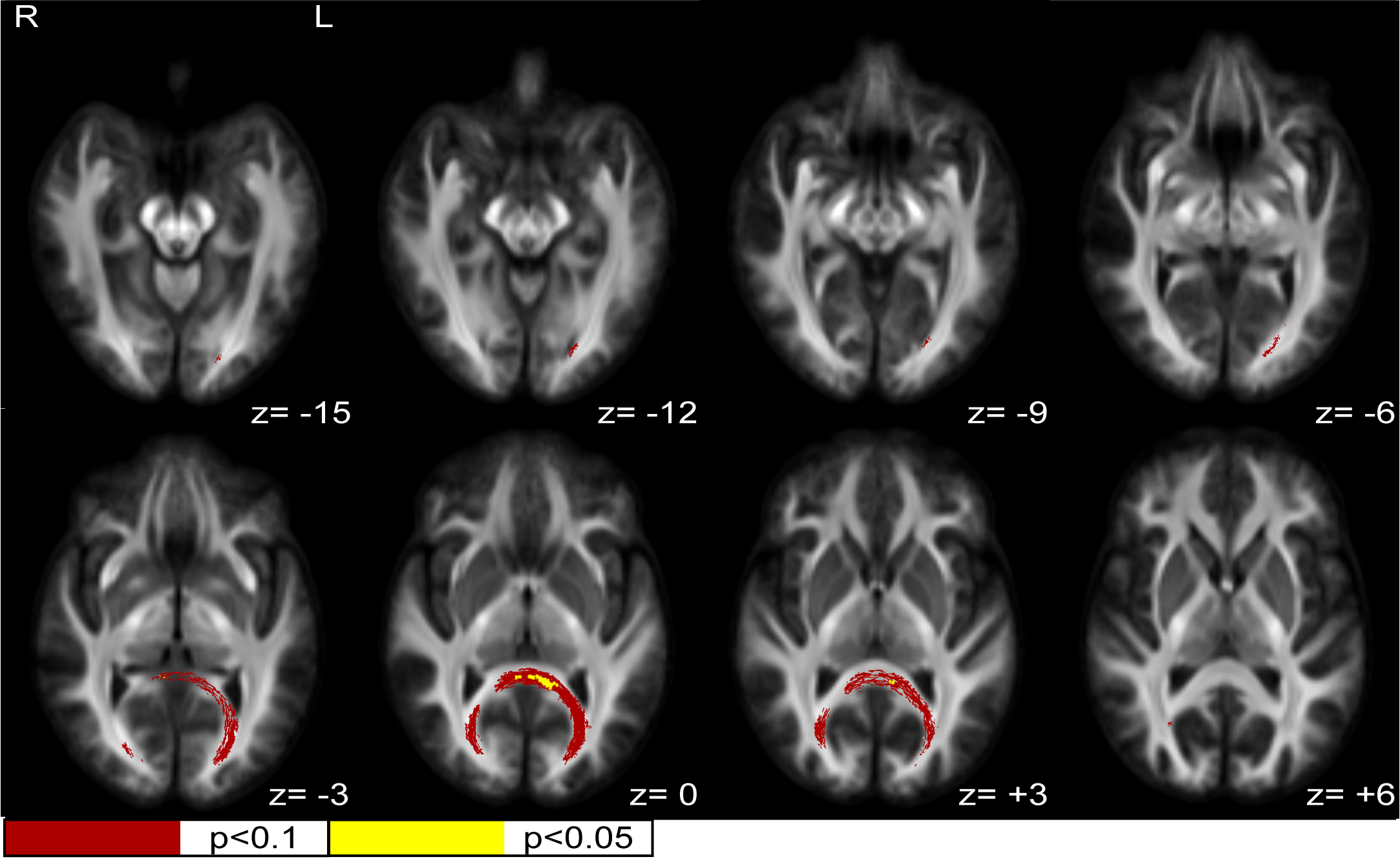
Figure 1. White matter fiber cross-section (FC) increases in neuropathic pain. Statistically significant (FWE-corrected) FC increases in SCI subjects with neuropathic pain compared to healthy SCI controls were seen in the posterior splenium of the corpus callosum/retrosplenial complex region. Z values are relative to the AC/PC plane.
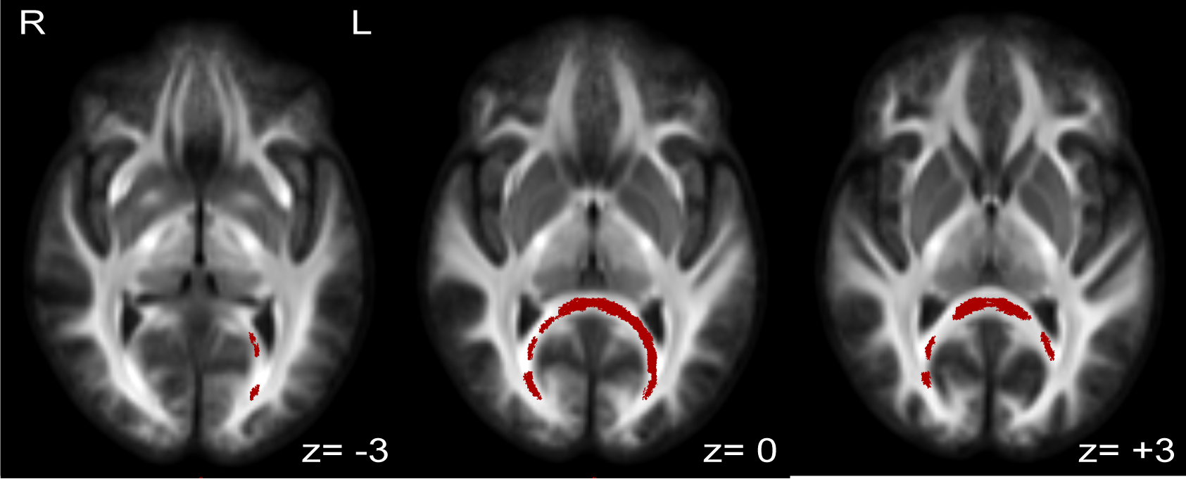
Figure 2. White matter fiber density and cross-section (FDC) increases in neuropathic pain. Statistically significant (FWE-corrected p<0.1) FDC increases in SCI subjects with neuropathic pain compared to healthy SCI controls were seen in the posterior splenium of the corpus callosum/ retrosplenial complex region. Z values are relative to the AC/PC plane.