XiaoQiang Du1, YunJing Xue1, HuaJun Chen1, and ZhongShuai Zhang2
1Fujian Medical University Union Hospital, Fuzhou, China, 2SIEMENS Healthcare, Shanghai, China
1Fujian Medical University Union Hospital, Fuzhou, China, 2SIEMENS Healthcare, Shanghai, China
We found WM abnormalities extending from
the motor to the extra-motor regions in ALS and observed a correlation between
distinct diffusion metrics and various clinical variables. In addition, LDH
generated the information that could complement conventional DTI metrics.
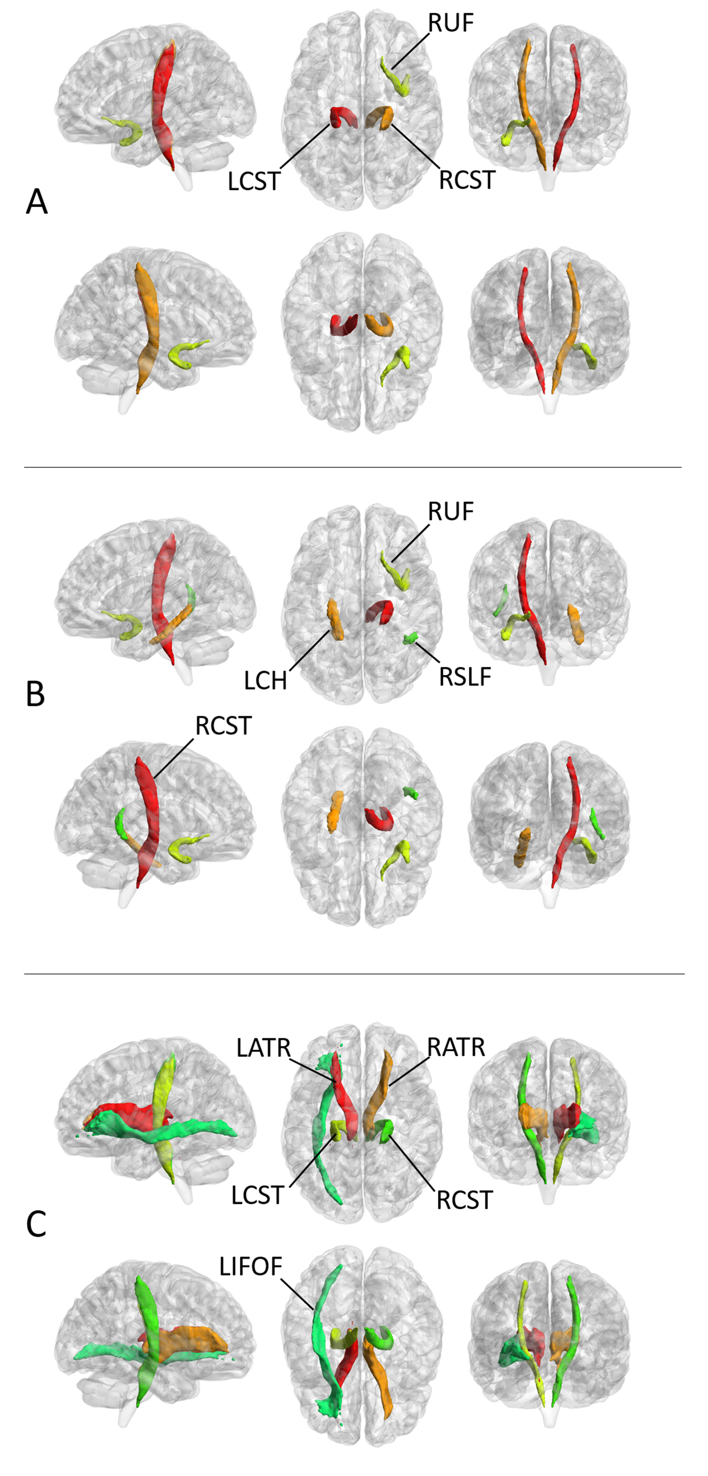
Fig. 2. The white matter (WM) tracts with
significant between-group differences in FA (A), MD (B) and LDH (C),
respectively. “R” and “L” indicate right and left side, respectively. RUF,
right uncinate fasciculus; RCST, right corticospinal tract; LCST, left
corticospinal tract; LCH, left cingulum hippocampus; RSLF, right superior
longitudinal fasciculus (temporal part); LATR, left anterior thalamic radiation;
RATR, right anterior thalamic radiation; LIFOF, left inferior frontal-occipital
fasciculus. These tracts were defined by the JHU ICBMDTI-81 white matter atlas.
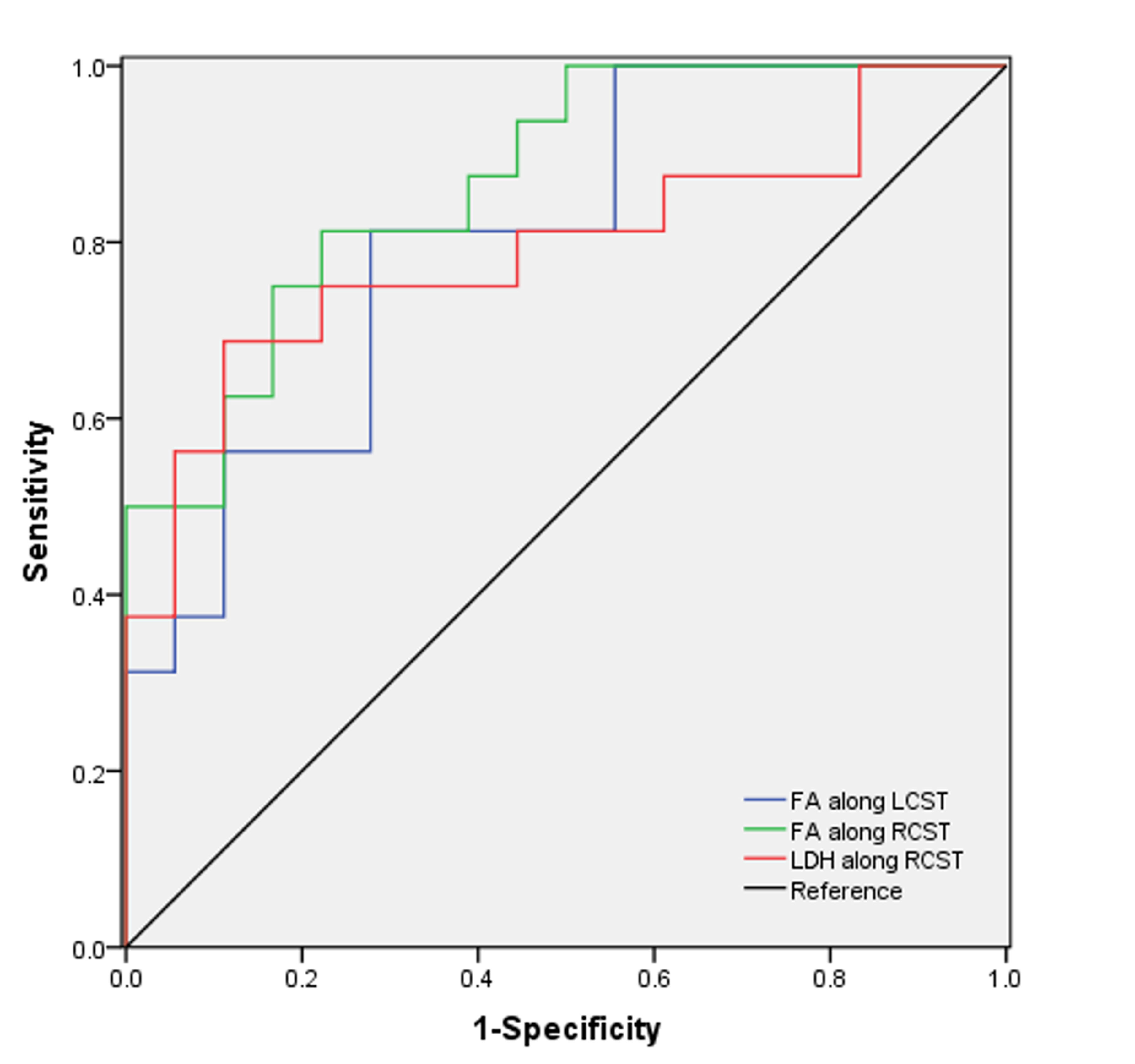
Fig. 4. The results of receiver operating characteristic (ROC) curve analysis.