Bhaswati Roy1, Sarah E Choi2, Milena Lai3, Luke Ehlert3, Rashmi Mullur4, Matthew J. Freeby4, and Rajesh Kumar3,5,6,7
1University of California at Los Angeles, LOS ANGELES, CA, United States, 2UCLA School of Nursing, University of California at Los Angeles, Los Angeles, CA, United States, 3Anesthesiology, University of California at Los Angeles, Los Angeles, CA, United States, 4Medicine, University of California at Los Angeles, Los Angeles, CA, United States, 5Radiology, University of California at Los Angeles, Los Angeles, CA, United States, 6Bioengineering, University of California at Los Angeles, Los Angeles, CA, United States, 7Brain Research Institute, University of California at Los Angeles, Los Angeles, CA, United States
1University of California at Los Angeles, LOS ANGELES, CA, United States, 2UCLA School of Nursing, University of California at Los Angeles, Los Angeles, CA, United States, 3Anesthesiology, University of California at Los Angeles, Los Angeles, CA, United States, 4Medicine, University of California at Los Angeles, Los Angeles, CA, United States, 5Radiology, University of California at Los Angeles, Los Angeles, CA, United States, 6Bioengineering, University of California at Los Angeles, Los Angeles, CA, United States, 7Brain Research Institute, University of California at Los Angeles, Los Angeles, CA, United States
Patients with Type 2 diabetes mellitus (T2DM) show brain tissue changes, but the nature and extent of damage and their progression with time are unclear. Using DTI based MD procedures, we showed chronic tissue changes in T2DM subjects and their continued progression after 6 months follow-up.
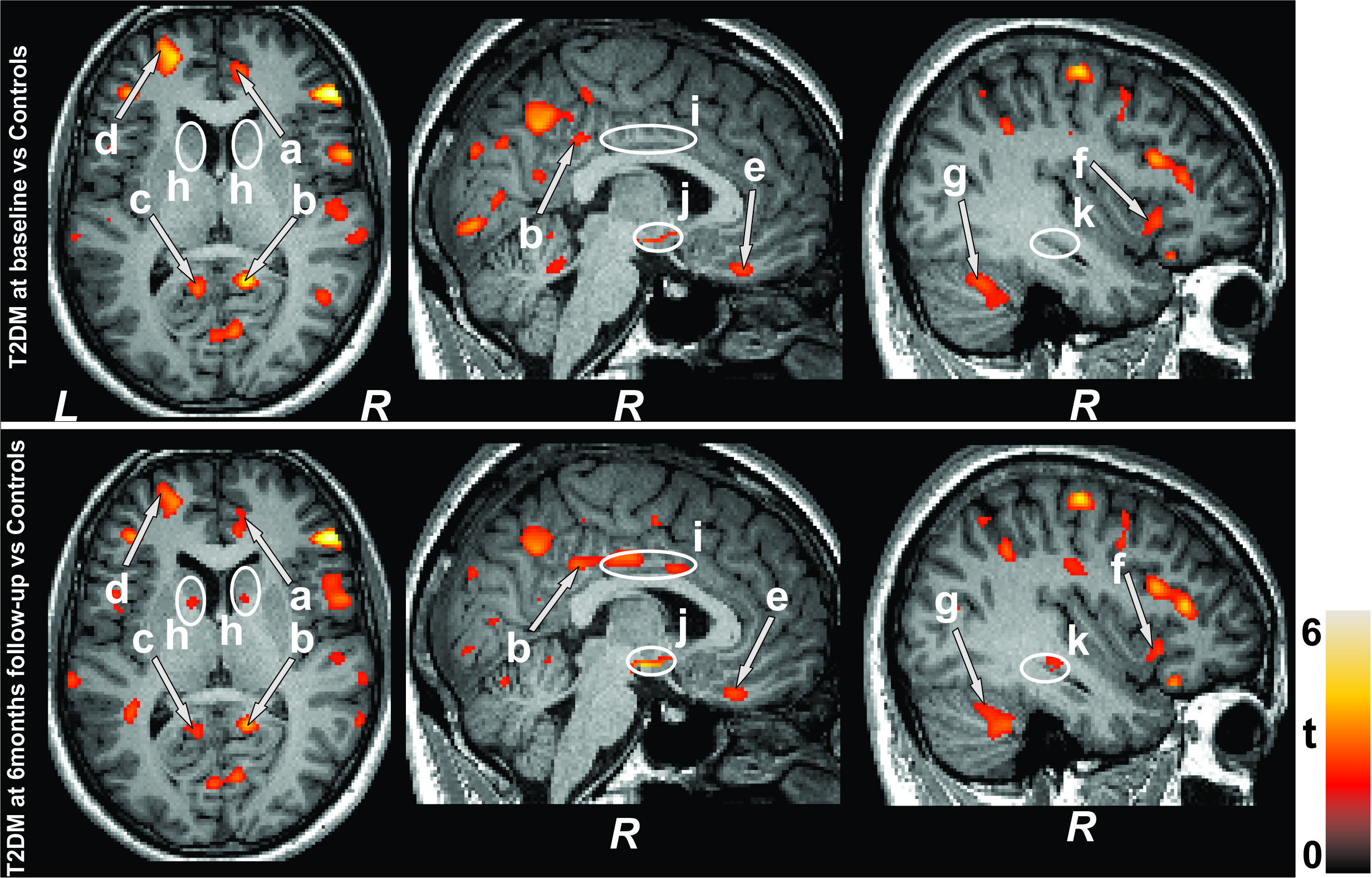
Figure 1: Brain regions with increased MD values in T2DM
patients at baseline and 6 months follow-up appeared over controls. These sites
included the (arrows) anterior and posterior cingulate (a-c), frontal
(d) and prefrontal (e), insular cortices (f), and cerebellum (g). Few
regions emerged with widespread damage in follow-up over baseline
T2DM patients, and these sites (ellipses) included the caudate (h),
middle cingulate (i), hypothalamus (j), and hippocampus (k). All images are in
neurological convention (L = left; R = right). Color bar indicates t-statistic
values.
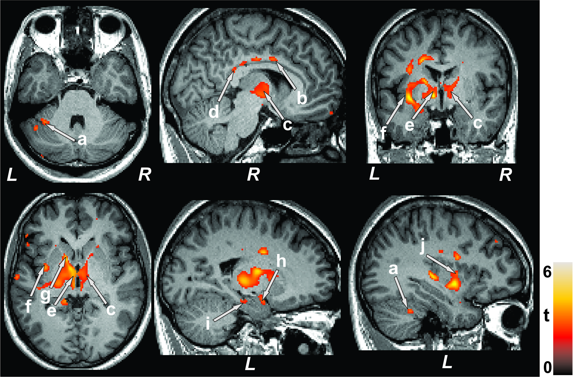
Figure 2: Paired t-test exhibit higher MD values in 6-months
follow-up T2DM patients over baseline. The regions with increased MD values
included the cerebellum (a), mid (b) and posterior (d) cingulate, thalamus (c,
e), insular cortices (f, j), caudate (g), amygdala (h), and hippocampus (i).
Figure conventions are same as in Figure 1.