Tiago Timoteo Fernandes1, Marc Golub1, Andreia Freitas1, Sairam Geethanath2,3, and Rita Gouveia Nunes1
1ISR, IST - University of Lisbon, Lisbon, Portugal, 2Medical Imaging Research Centre, Dayananda Sagar Institutions, Bangalore, India, 3Magnetic Resonance Research Center, Columbia University, NY, DC, United States
1ISR, IST - University of Lisbon, Lisbon, Portugal, 2Medical Imaging Research Centre, Dayananda Sagar Institutions, Bangalore, India, 3Magnetic Resonance Research Center, Columbia University, NY, DC, United States
Diffusion-weighted Imaging (DWI) can detect ischemic tissue in stroke patients. Ultra-low field systems (ULF) could provide a point-of-care solution but suffer from low SNR. This simulation work investigates the feasibility of DWI at ULF.
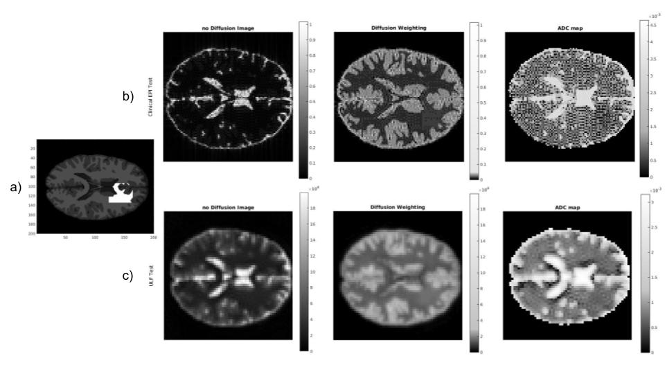
Figure 4 - Noise free simulations for two different sequences and their reconstructed images. a) Sample with four different tissues each with their respective relaxation times. The LS tissue (in white) has similar values to the WM. b) Partial EPI sequence in clinical MR scheme with b=1000 s/mm2, B0=1.5T, sm=200T/m/s, gm=30mT/m, res=2mm. c) Spiral Sequence, single shot in LF with b=700 s/mm2, B0=0.2T, sm=50T/m/s, gm =14mT/m, res=3mm. Images were reconstructed for both sequence with no DW (left), with DW (same color bar than no DW) (center) and ADC maps (right).
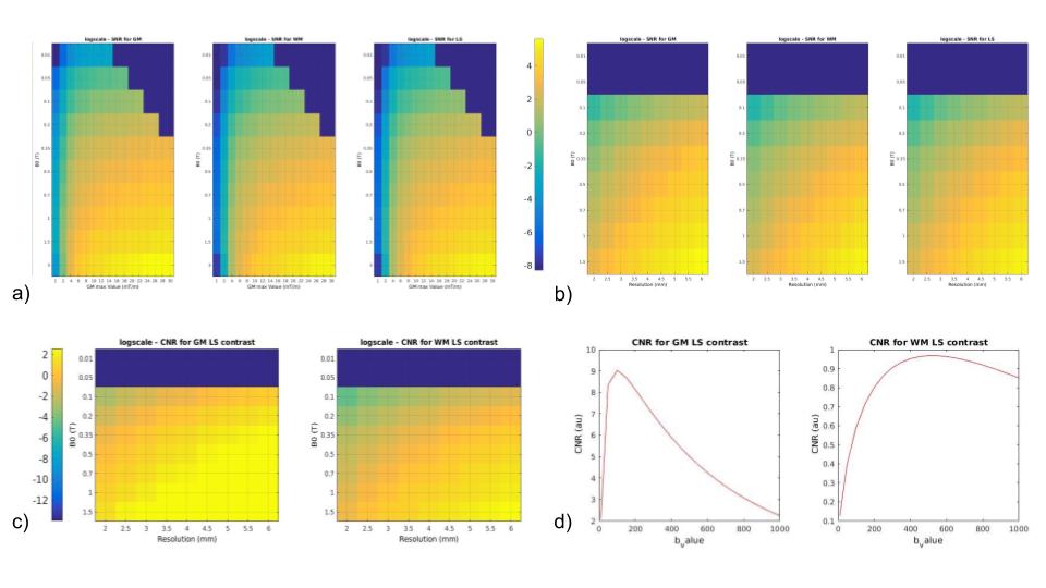
Figure 2 - SNR and CNR for varying B0 field intensity, gm, spatial resolution (displayed using a logarithmic scale) and b-values. a) Variation of SNR across gm values and B0 field, for a fixed res = 4mm. b) Variation of SNR across resolution values and B0 field, for a fixed gm = 20 mT/m. c) Variation of CNR across resolution values and B0 field, for a fixed gm = 20 mT/m. d) Variation of CNR across b-values, for a fixed gm = 20 mT/m and res = 4mm.