Jin Gao1,2, Zachery Morrissey3, Alex Leow3, Orly Lazarov4, Danilo Erricolo1, Richard Magin5, and Weiguo Li2,5
1Electrical and Computer Engineering, University of Illinois at Chicago, Chicago, IL, United States, 2Research Resources Center, University of Illinois at Chicago, Chicago, IL, United States, 3Department of Psychiatry, University of Illinois at Chicago, Chicago, IL, United States, 4Department of Anatomy and Cell Biology, University of Illinois at Chicago, Chicago, IL, United States, 5Department of Bioengineering, University of Illinois at Chicago, Chicago, IL, United States
1Electrical and Computer Engineering, University of Illinois at Chicago, Chicago, IL, United States, 2Research Resources Center, University of Illinois at Chicago, Chicago, IL, United States, 3Department of Psychiatry, University of Illinois at Chicago, Chicago, IL, United States, 4Department of Anatomy and Cell Biology, University of Illinois at Chicago, Chicago, IL, United States, 5Department of Bioengineering, University of Illinois at Chicago, Chicago, IL, United States
Diffusion MRI offers unique insights into the pathophysiology of AD in vivo. This feasibility study aims to assess and visualize the white matter changes using an ultrahigh b-value diffusion MRI.
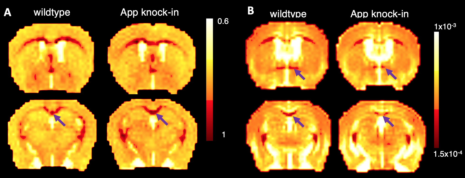
Fig. 3. Parameters maps generated from CTRW
diffusion model for two image slices of an APP knock-in mouse and corresponding
slices in WT control. A. α map with arrow pointed to CC; B, D maps (mm2/s)
with arrow
pointed to AC (upper panel) and CC (lower panel).
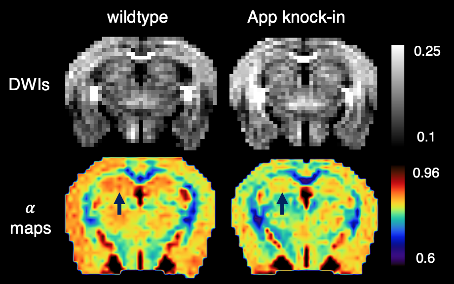
Fig. 4. Hippocampus showing lower values in α maps (lower panel) in APP knock-in mouse
comparing to the WT control. Upper panel: diffusion-weighted images
with b = 4050 s/mm2,
with gray bar showing the normalized signal intensity.
Lower panel: α maps with color bar showing the
extracted α values. Arrow points to the hippocampus.