Álvaro Planchuelo-Gómez1, Santiago Aja-Fernández1, David García-Azorín2, Ángel Luis Guerrero2, and Rodrigo de Luis-García1
1Imaging Processing Laboratory, Universidad de Valladolid, Valladolid, Spain, 2Headache Unit, Hospital Clínico Universitario de Valladolid, Valladolid, Spain
1Imaging Processing Laboratory, Universidad de Valladolid, Valladolid, Spain, 2Headache Unit, Hospital Clínico Universitario de Valladolid, Valladolid, Spain
Diffusion MRI scalar measures are more precise
employing 61 gradient directions in comparison to 40 and 21 directions. Equivalent
results to the 61 gradient directions case can be obtained with 40 and 21
directions by increasing the sample size.
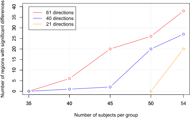
Figure 2. Evolution in the number of regions with significant
differences using 61, 40 and 21 diffusion gradient directions. Axial diffusivity was compared between chronic and
episodic migraine patients. A decrease in the number of gradients can be compensated by increasing sample size. Number of subjects per group for the diverse iterations is shown in the horizontal axis, corresponding the maximum value to
the original sample. Median values are used as punctual estimations. Red, blue
and orange represent the number of regions using 61, 40 and 21 gradient
directions respectively.
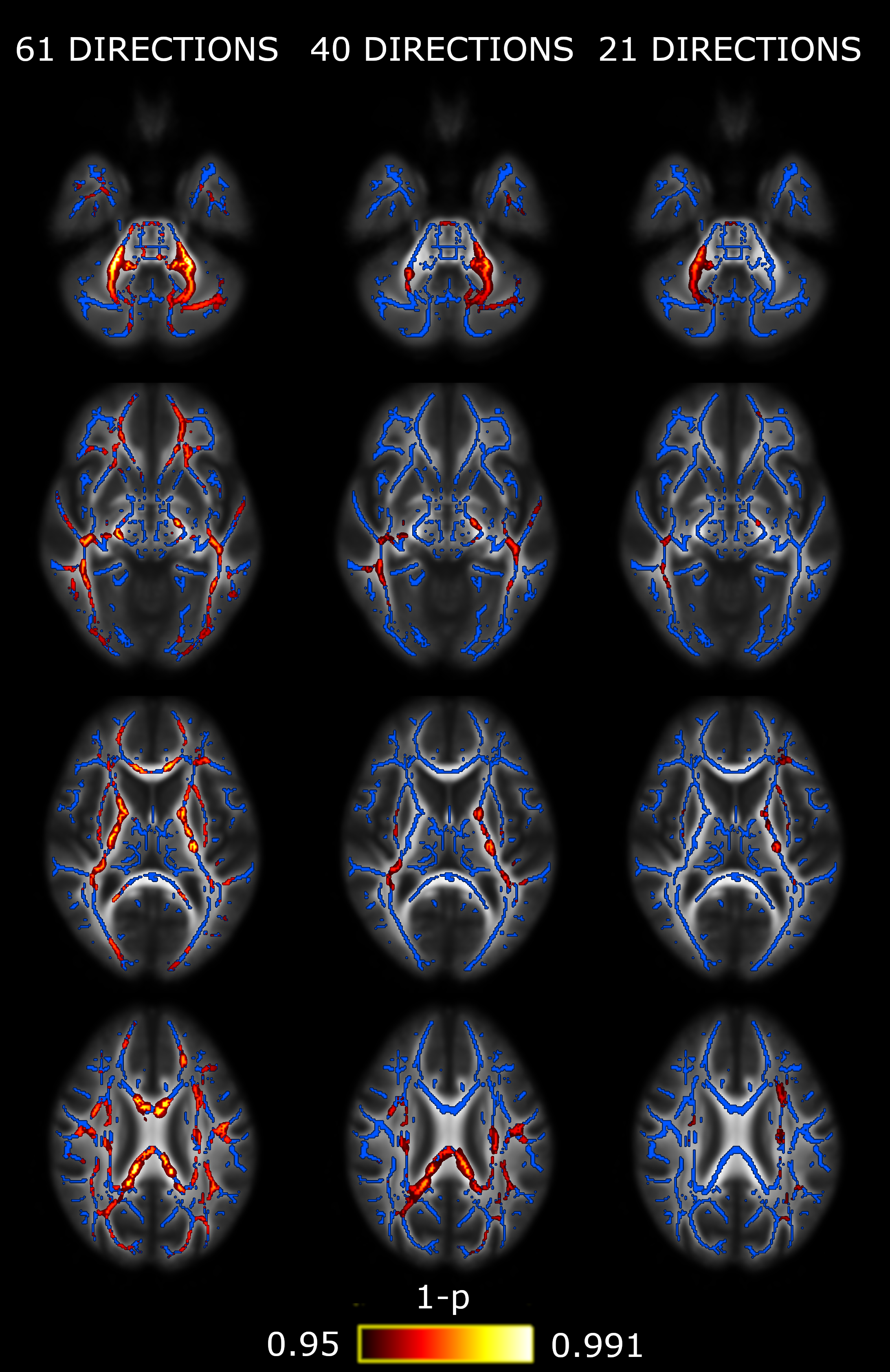
Figure 1. Visual comparison of the results with the
diverse diffusion encoding schemes. Results with the original sample comparing axial diffusivity between
chronic and episodic migraine patients are shown. The first column contains the
results for 61 gradient directions (37 regions with significant differences), and the second and third for 40 (27 regions) and 21
directions (20 regions) respectively. White matter skeleton is shown in blue and voxels with
significant differences in red-yellow. The color bar shows the 1-p values
(family-wise error corrected).