Huiqin Zhang1, Hui Zhang1, Franki Kai-Hei Tse 2, Edward Sai-Kam Hui1, Peng Cao1, Kannie Wai Yan Chan3, Queenie Chan4, Karl HERRUP5, and Henry Ka Fung Mak1
1Department of Diagnostic Radiology, The University of Hong Kong, Hong Kong, Hong Kong, 2Department of Health Technology and Informatics, The Hong Kong Polytechnic University, Hong Kong, Hong Kong, 3Department of Biomedical Engineering, City University of Hong Kong, Hong Kong, Hong Kong, 4Philips Healthcare, Hong Kong, Hong Kong, 5Department of Neurobiology, University of Pittsburgh, Pittsburgh, PA, United States
1Department of Diagnostic Radiology, The University of Hong Kong, Hong Kong, Hong Kong, 2Department of Health Technology and Informatics, The Hong Kong Polytechnic University, Hong Kong, Hong Kong, 3Department of Biomedical Engineering, City University of Hong Kong, Hong Kong, Hong Kong, 4Philips Healthcare, Hong Kong, Hong Kong, 5Department of Neurobiology, University of Pittsburgh, Pittsburgh, PA, United States
AD appears
to have similar microstructural WM changes as RRMS, which might indicate that
they share similar pathogenetic mechanisms such as demyelination.
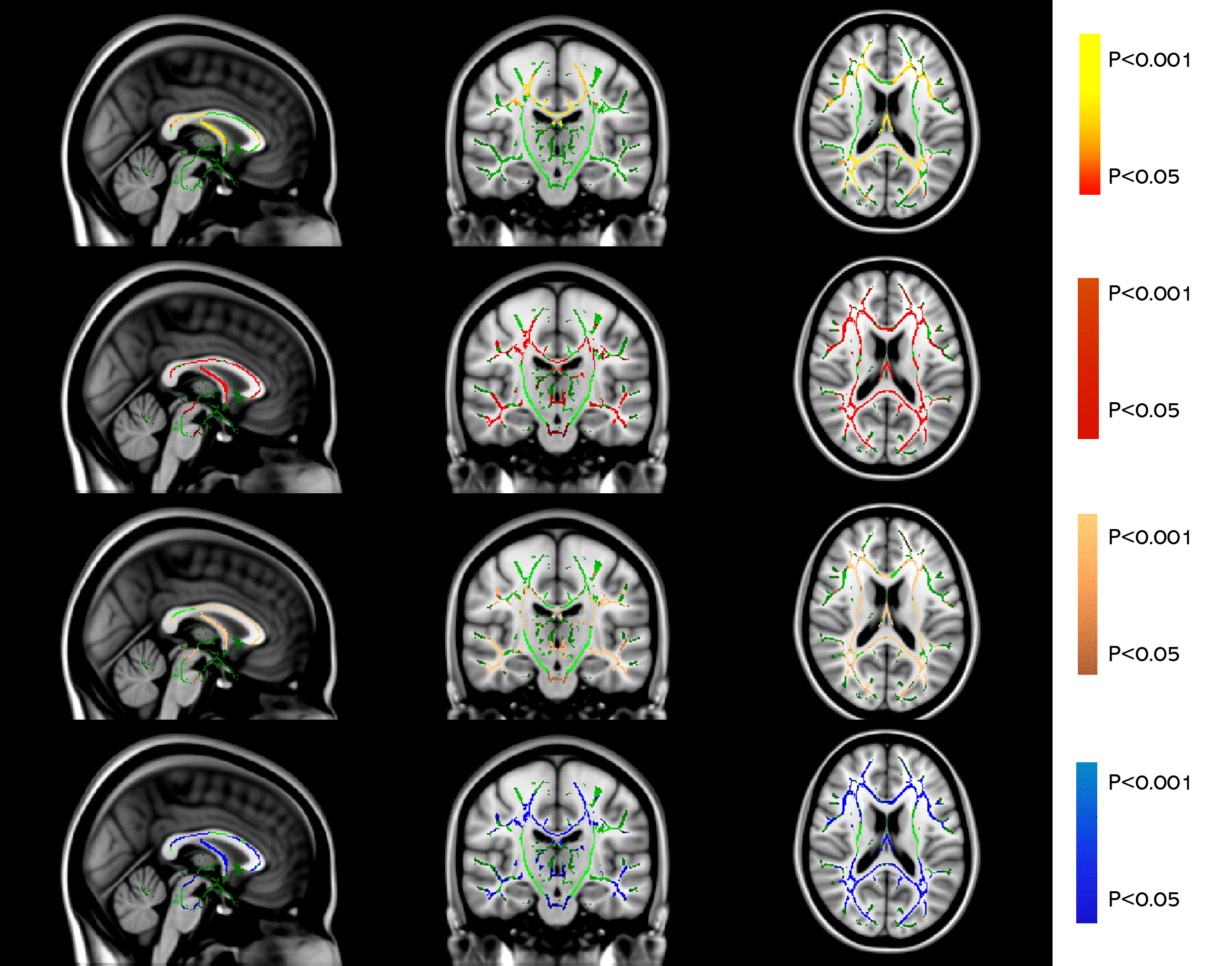
Fig 1a. TBSS results of FA, MD, AxD and RD images
between normal controls and AD patients. Green represents mean FA skeleton of all
participants; yellow-red, red, copper, and blue represent regions with
decreased FA (1st row), increased MD (2nd row), increased AD (3rd row) and
increased RD (4th row) separately in AD patients (P < 0.05, TFCE corrected
for multiple comparisons)
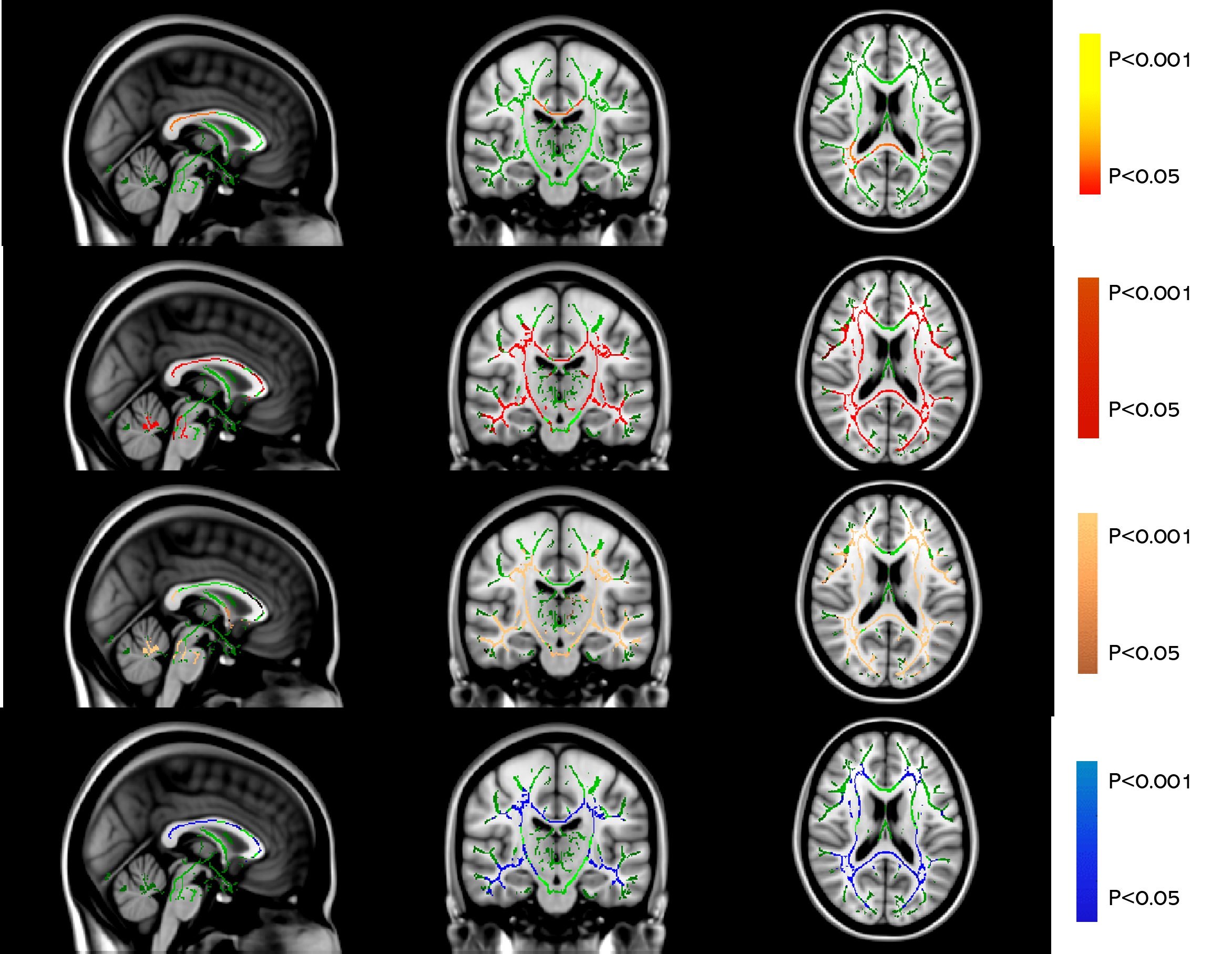
Fig 1b. TBSS results of FA, MD, AxD and RD images
between normal controls and MS patients. Green
represents mean FA skeleton of all participants; yellow-red, red, copper, and blue
represent regions with decreased FA (1st row), increased MD (2nd row),
increased AD (3rd row) and increased RD (4th row) separately in MS patients (P
< 0.05, TFCE corrected for multiple comparisons).