Ethan A Cook1, Shannon E Callen2, Shilpa Buch2, and Balasrinivasa R Sajja3
1College of Medicine, University of Nebraska Medical Center, Omaha, NE, United States, 2Pharmacology and Experimental Neuroscience, University of Nebraska Medical Center, Omaha, NE, United States, 3Radiology, University of Nebraska Medical Center, Omaha, NE, United States
1College of Medicine, University of Nebraska Medical Center, Omaha, NE, United States, 2Pharmacology and Experimental Neuroscience, University of Nebraska Medical Center, Omaha, NE, United States, 3Radiology, University of Nebraska Medical Center, Omaha, NE, United States
In vivo DTI detected brain structural changes in cocaine administered mice. Our findings indicated demyelination in white matter structures in chronic administration in mice. Results also indicated the involvement of gray matter.
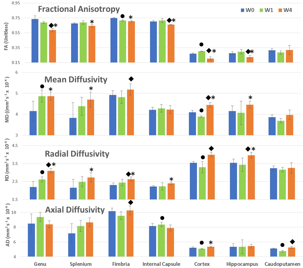
Figure 3: Change in DT measures at different time points. Blue, green, and orange
colors denote values at week 0, week 1, and week 4 respectively. Statistically
significant change is represented by symbol as ●: (W0~W1), ◆: (W1~W4), *: (W0~W4).
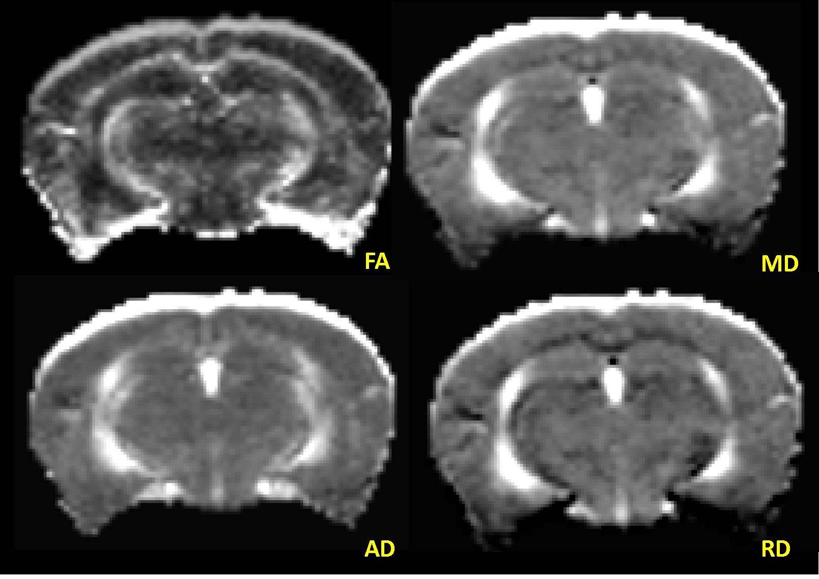
Figure 1: Representative
FA, MD, AD, and RD maps from same cross-section of a mouse brain.