Bing Yao1,2, Hannah Ovadia1, Gail Forrest3, and Steven Kirshblum2,4
1Rocco Ortenzio Neuroimaging Center, Kessler Foundation, West Orange, NJ, United States, 2Department of Physical Medicine and Rehabilitation, Rutgers University, Newark, NJ, United States, 3Center for Mobility and Rehabilitation Engineering Research, Kessler Foundation, West Orange, NJ, United States, 4Kessler Institute for Rehabilitation, West Orange, NJ, United States
1Rocco Ortenzio Neuroimaging Center, Kessler Foundation, West Orange, NJ, United States, 2Department of Physical Medicine and Rehabilitation, Rutgers University, Newark, NJ, United States, 3Center for Mobility and Rehabilitation Engineering Research, Kessler Foundation, West Orange, NJ, United States, 4Kessler Institute for Rehabilitation, West Orange, NJ, United States
Our study demonstrates that DTI may serve as a
tool to assess the changes at different regions of the spine on spinal cord injured patients during their six months recovery period after injury, of which
information is usually hard to be obtained by traditional evaluation methods.
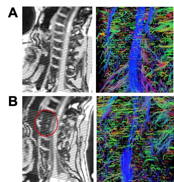
Fig. 1: Comparison of the spine MR images and tractography
for a SCI patient (B) and a matched healthy control (A). The red circle indicates a
significant signal dropout at the C4 level, which matches the location of the
implanted hardware in this SCI patient.
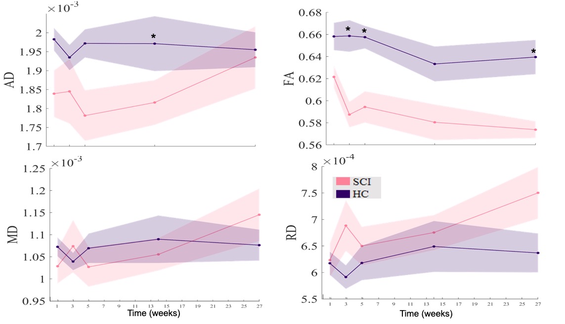
Fig. 3: Above Injury Level. Mean
values of AD, FA, MD, and RD above the area of injury across five visits. Values represent mean ± SEM. There were significant differences in AD and FA between groups,
indicating by “*” (p < 0.05).
