Zhang Jiulong1, Shi Nannan1, Zhang Yijun1, Ye Wen1, Zhang yong2, Shan fei1, and Shi Yuxin1
1radiology department, Shanghai public health clinical center, shanghai, China, 2General surgery, Shanghai public health clinical center, shanghai, China
1radiology department, Shanghai public health clinical center, shanghai, China, 2General surgery, Shanghai public health clinical center, shanghai, China
The quantitative parameters of IVIM-DWI combined with sDKI models are helpful to improve the accuracy and specificity in differential diagnosis of RPLS subtypes and have the potentiality to predict the histological grading of RPLS.
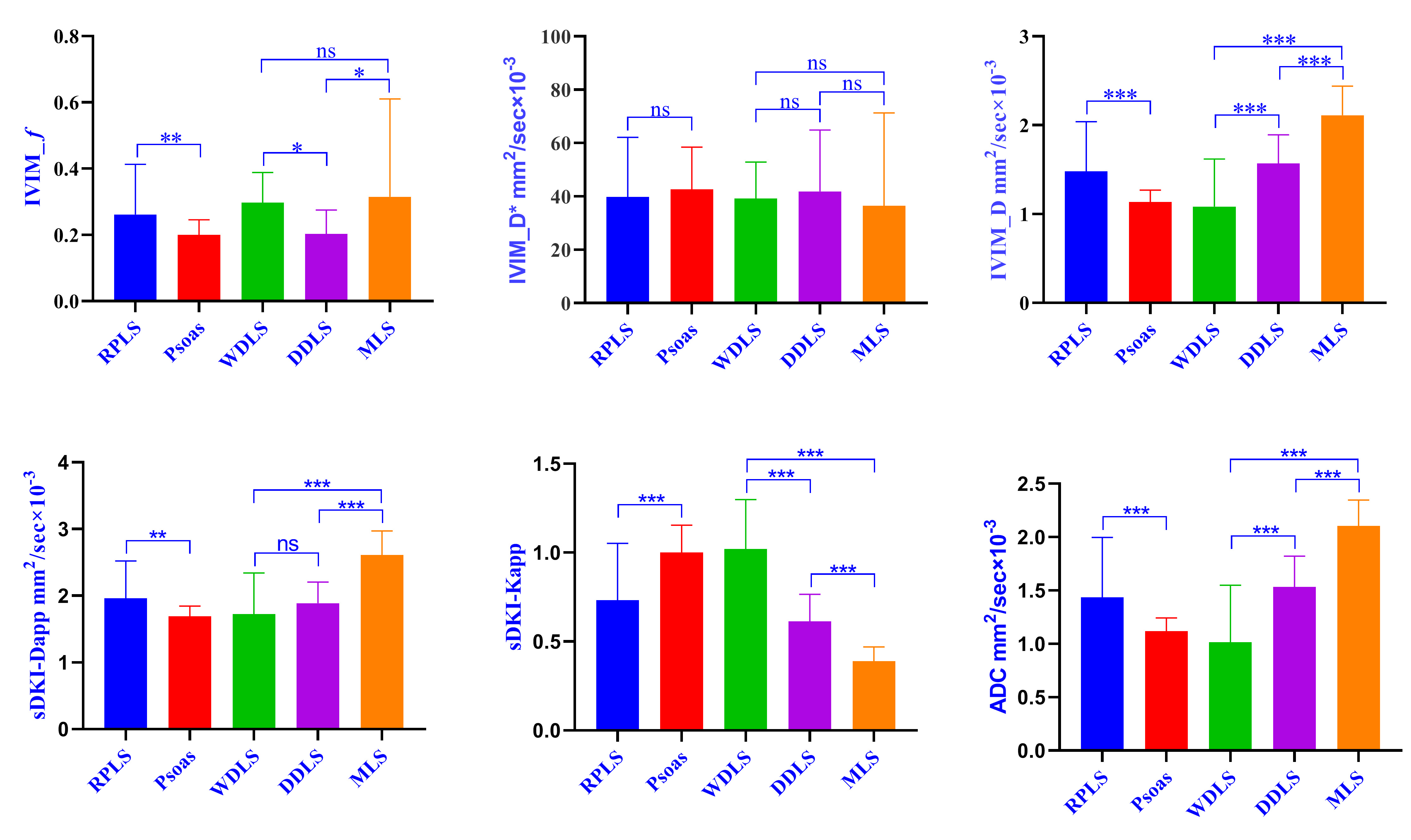
Fig.1. Column bar graph (Mean with SD) of the parameters (f, D*, D, Dapp, Kapp and ADC) of IVIM-DWI and sDKI models in psoas group, retroperitoneal liposarcoma (RPLS) and its subgroups (well-differentiated liposarcoma (WDLS), dedifferentiated liposarcoma (DDLS), myxoid liposarcoma (MLS)), which were analyzed by LSD test. (ns p>0.05, * p<0.05, ** p<0.01, *** p<0.001)
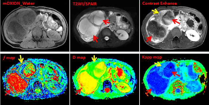
A 34-year-old woman with a large retroperitoneal soft tissue mass. mDXION-water phase showed iso-signal or low signal, while T2WI/SPAIR image showed high signal. There were more mucous components in the lesion, and the solid components were obviously enhanced after enhancement, while the mucous area showed uneven flocculent enhancement. IVIM-DWI and sDKI parameter maps: mean f value=62%, mean D value=2.02 mm2/sec×10-3, mean Kapp value=0.33. Postoperative pathology confirmed MLS.