Shao-Chieh Lin1, Jui-Heng Lin1, Chun-Jung Juan2,3,4, Kai-Min Chien5, Teng-Yi Huang6, Yi-Jui Liu7, Chang Hsien Liu 5, Ya-Hui Li5, Szu Hsien Chou 5, and Chi-Feng Hsieh5
1Master 's Program of Biomedical Informatics and Biomedical Engineering, Feng Chia University, Taichung, Taiwan, 2Department of Medical Imaging, China Medical University Hsinchu Hospital, Hsinchu, Taiwan, 3Department of Radiology, School of Medicine, College of Medicine, China Medical University, Taichung, Taiwan, 4Department of Computer Science and Information Engineering, National Taiwan University, Taipei, Taiwan, 5Department of Medical Imaging, Chinese Medical University Hsinchu Hospital, Hsinchu, Taiwan, 6Department of Electrical Engineering, National Taiwan University of Science and Technology, Taipei, Taiwan, 7Department of Automatic Control Engineering, Feng Chia University, Taichung, Taiwan
1Master 's Program of Biomedical Informatics and Biomedical Engineering, Feng Chia University, Taichung, Taiwan, 2Department of Medical Imaging, China Medical University Hsinchu Hospital, Hsinchu, Taiwan, 3Department of Radiology, School of Medicine, College of Medicine, China Medical University, Taichung, Taiwan, 4Department of Computer Science and Information Engineering, National Taiwan University, Taipei, Taiwan, 5Department of Medical Imaging, Chinese Medical University Hsinchu Hospital, Hsinchu, Taiwan, 6Department of Electrical Engineering, National Taiwan University of Science and Technology, Taipei, Taiwan, 7Department of Automatic Control Engineering, Feng Chia University, Taichung, Taiwan
ADC measurement in the slice with the largest tumor which tumor size over one third of the total tumor volume, could instead the ADC measurement of whole tumor for diagnosis of the PMA, WT and MT in parotid gland.
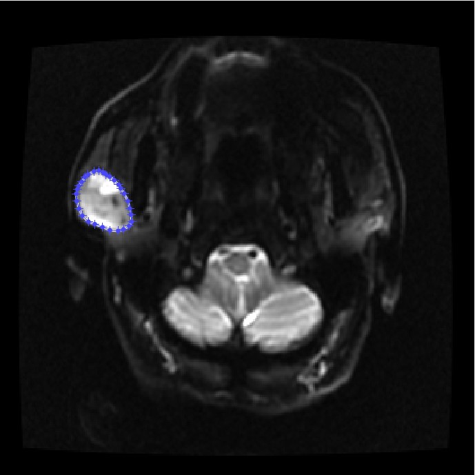
Figure 1. Illustration of manual contouring
of a parotid tumor (blue) on EP-DWI (b0) for further calculating the mean and standard
derivation on ADC map.
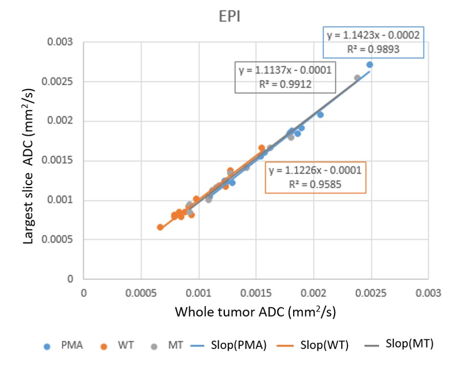
Figure
2. ADC of parotid gland tumors of PMA, WT and MT on the largest slice and whole
tumor. PMA: pleomorphic adenoma; WT: Warthin’s tumor; *** represents a P <0.001.