Kurt G Schilling1, Kristin P O'Grady1,2, Samantha By3, Haley Feiler1, Francesca Bagnato4, Bennett A Landman1,5,6,7, and Seth A Smith1,5,8
1Vanderbilt University Institute of Imaging Science, Nashville, TN, United States, 2Radiology and Radiological Sciences, Vanderbilt University Medial Center, Nashvillet, TN, United States, 3Hyperfine Research Inc, Guilford, CT, United States, 4Neurology, Vanderbilt University Medical Center, Nashville, TN, United States, 5Biomedical Engineering, Vanderbilt University, Nashville, TN, United States, 6Radiology and Radiological Sciences, Vanderbilt University Medical Center, Nashville, TN, United States, 7Electrical Engineering, Vanderbilt University, Nashville, TN, United States, 8Radiology and Radiological Sciences, Vanderbilt University, Nashville, TN, United States
1Vanderbilt University Institute of Imaging Science, Nashville, TN, United States, 2Radiology and Radiological Sciences, Vanderbilt University Medial Center, Nashvillet, TN, United States, 3Hyperfine Research Inc, Guilford, CT, United States, 4Neurology, Vanderbilt University Medical Center, Nashville, TN, United States, 5Biomedical Engineering, Vanderbilt University, Nashville, TN, United States, 6Radiology and Radiological Sciences, Vanderbilt University Medical Center, Nashville, TN, United States, 7Electrical Engineering, Vanderbilt University, Nashville, TN, United States, 8Radiology and Radiological Sciences, Vanderbilt University, Nashville, TN, United States
We investigate multi-compartment diffusion MRI models in the in vivo spinal cord of MS patients. We find differences in diffusion measures between patients and controls, and characterize relationships of indices with clinical disability measures.
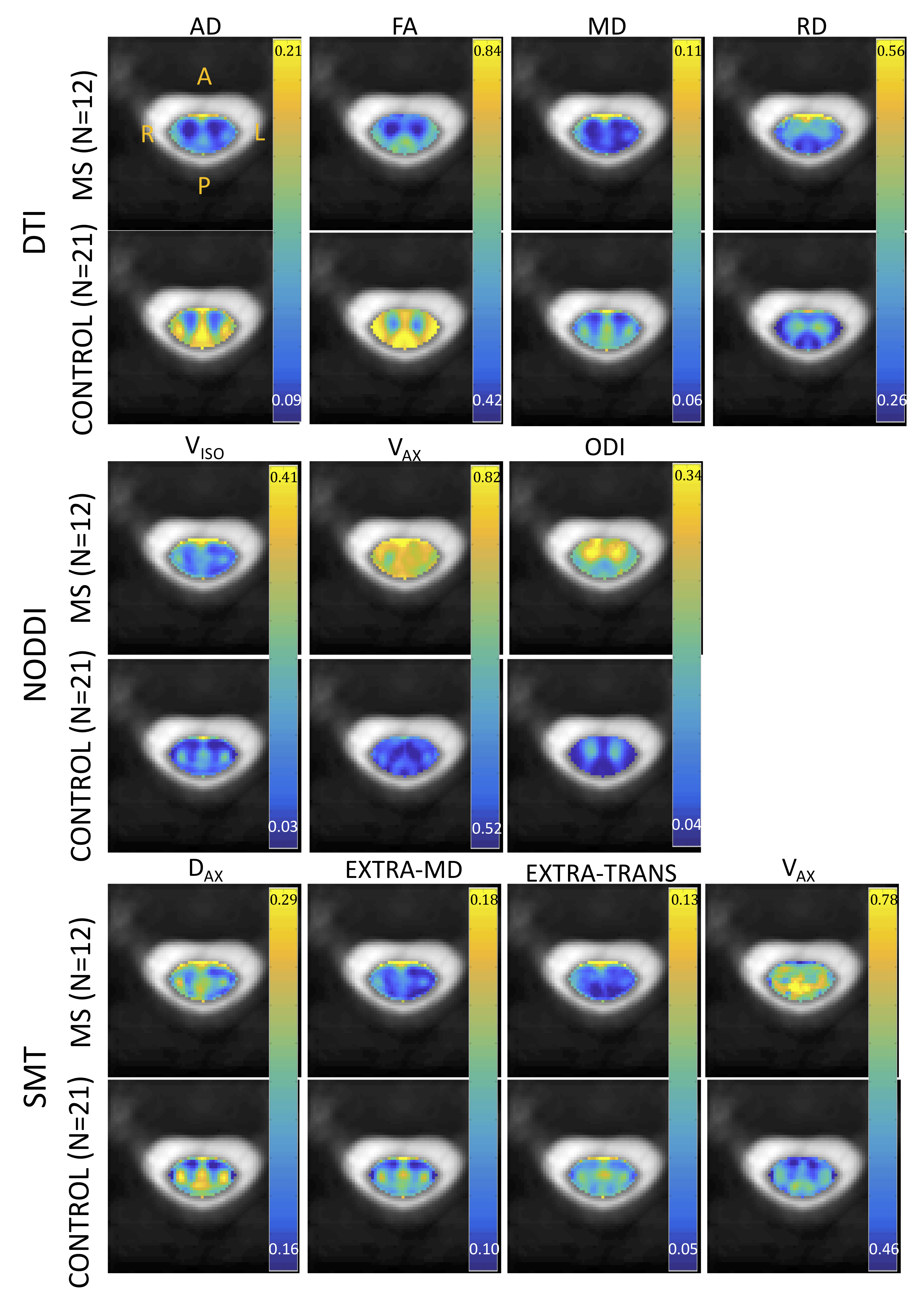
Figure 1. Qualitative differences are apparent in multi-compartment indices between controls (N=21) and MS patients (N=12). Indices are registered to template space and averaged. For DTI (top), NODDI (middle), and SMT (bottom), indices for control and MS are displayed on the same scale.
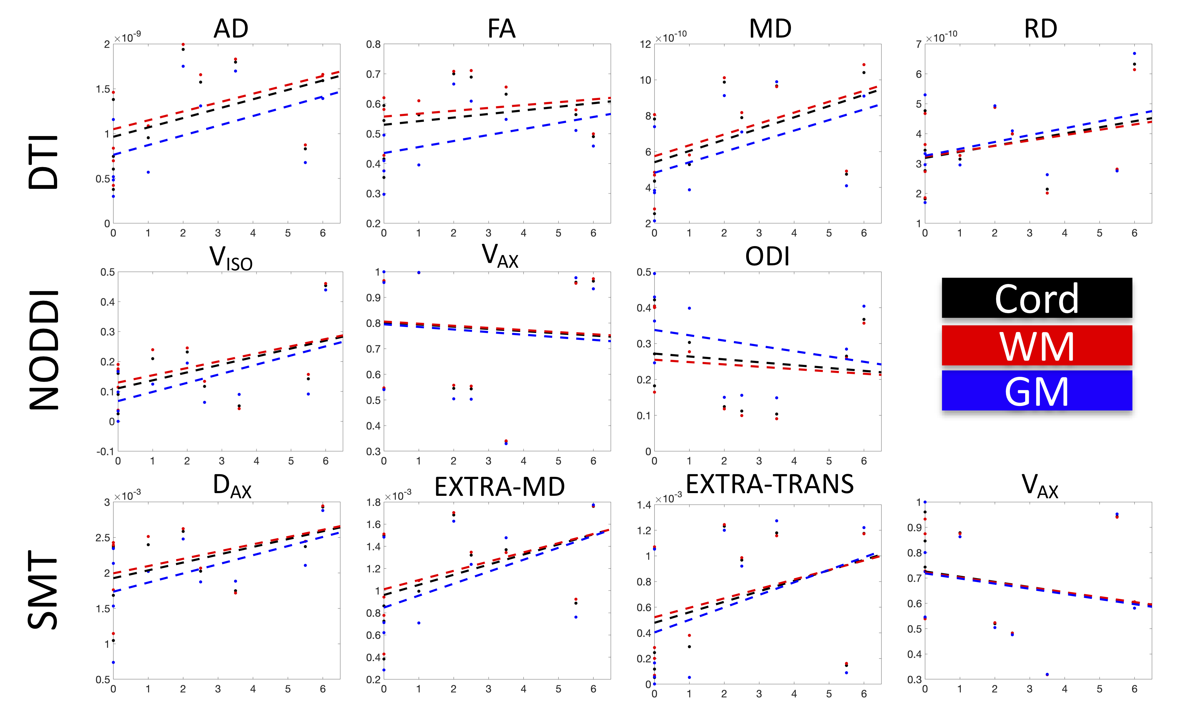
Figure 3. Multi-compartment indices derived from DTI (top), NODDI (middle), and SMT (bottom) are plotted against EDSS score for all MS patients (Note that 10 of the 12 MS patients had clinical EDSS scores). Lines of best fit are shown over the whole cord (black), and in WM (red) and GM (blue) regions. No correlations were statistically significant – although clear (nonlinear) trends are apparent in many indices.