Dimitra Flouri1,2, Jack RT Darby3, Stacey L Holman3, Sunthara R Perumal4, Anna L David5,6, Andrew Melbourne1,2, and Janna L Morrison3
1School of Biomedical Engineering & Imaging Sciences, King's College London, London, United Kingdom, 2Department of Medical Physics & Biomedical Engineering, University College London, London, United Kingdom, 3Early Origins of Adult Health Research Group, University of South Australia, Adelaide, Australia, 4Preclinical Imaging and Research Laboratories, South Australian Health and Medical Research Institute, Adelaide, Australia, 5Institute for Women's Health, University College London, London, United Kingdom, 6NIHR University College London Hospitals Biomedical Research Center, London, United Kingdom
1School of Biomedical Engineering & Imaging Sciences, King's College London, London, United Kingdom, 2Department of Medical Physics & Biomedical Engineering, University College London, London, United Kingdom, 3Early Origins of Adult Health Research Group, University of South Australia, Adelaide, Australia, 4Preclinical Imaging and Research Laboratories, South Australian Health and Medical Research Institute, Adelaide, Australia, 5Institute for Women's Health, University College London, London, United Kingdom, 6NIHR University College London Hospitals Biomedical Research Center, London, United Kingdom
This study characterises diffusion and perfusion properties
of the placenta such as the apparent diffusion coefficient, T2 measurements,
fractional anisotropy and perfusion fraction derived from IVIM-analysis on sheep placental to validate new imaging markers
of placental function.
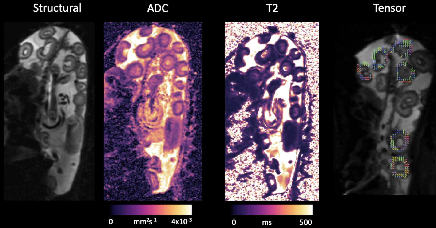
Figure 1: Figure shows example of the ADC, T2
and FA maps of a single subject.
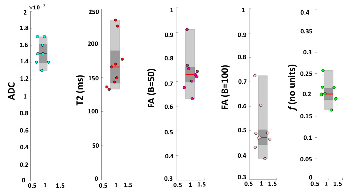
Figure 3:
Box-plots summarising results over the 9 singleton pregnancies. Each plot
shows: the median (red line), the 25th and 75th
percentile (grey box) and individual means of each subject ROIs (circles).