Junting Zou1, Jilei Zhang2, Yuanyuan Xie3, yunpeng Shen4, Jiacheng Du4, Yang Chen4, Yang Chen4, Bing Zhang3, and Xiaoli Mai5
1Nanjing Drum Tower Hospital Clinical College of Nanjing Medical University, Nanjing, China, 2Clinical Science, Philips Healthcare, Shanghai, China, 3Nanjing Drum Tower Hospital, Nanjing, China, 4Southeast University, Nanjing, China, 5Radiology, Nanjing Drum Tower Hospital, Nanjing, China
1Nanjing Drum Tower Hospital Clinical College of Nanjing Medical University, Nanjing, China, 2Clinical Science, Philips Healthcare, Shanghai, China, 3Nanjing Drum Tower Hospital, Nanjing, China, 4Southeast University, Nanjing, China, 5Radiology, Nanjing Drum Tower Hospital, Nanjing, China
The DTI can be of great value in the dynamic evaluation of morphological and neurological changes in beagles after TSCI. The FA value can satisfactorily evaluate the completeness of the fiber tracts after SCI and the repair of them by stem cells.
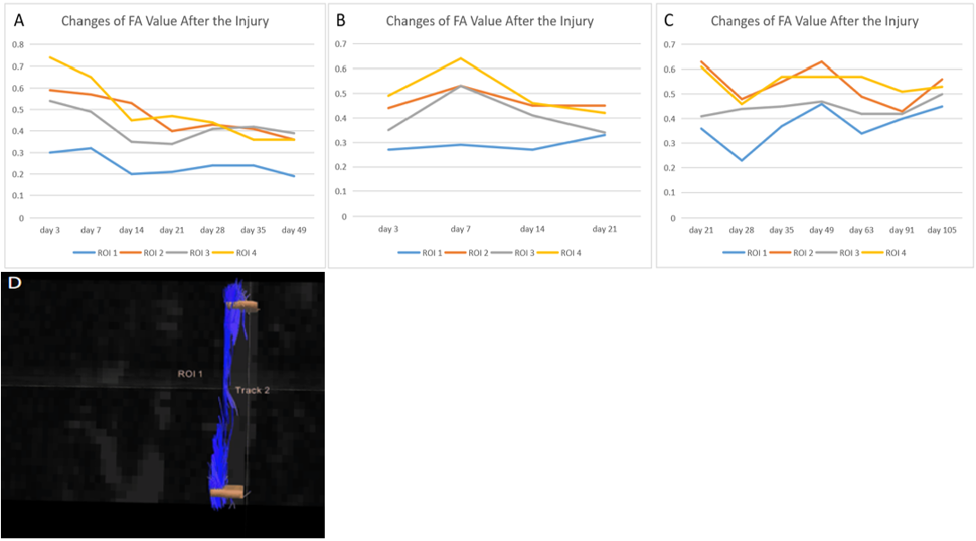
Figure 4: Dynamic changes of FA value after TSCI. ROI 1 to 4 represent injured and uninjured side of the injury site and rostral to the injury site. A. Group C: The FA value of all ROIs decreases with time, probably for limited self-repair ability to recover from SCI. B. Group T: Changes of FA value in the three uninjured areas are consistent and is higher than that in the injured area, where it increases with time. C. Group M: The FA value of the injured area increases with time, suggesting that MSCs may repair injured fiber tracts. D. The DTT modeling can observe that the fiber tracts are broken.
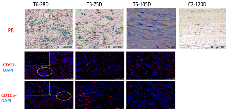
Figure 5: Pathological and immunohistochemical examination results. T6-28d: CD90+CD105 immunofluorescent staining confirms that there exists SPIO in living hUC-MSCs. T3-75d: CD90+CD105 immunofluorescent staining finds that MSC presents abnormal living cell morphology. It can be speculated that transplanted MSCs have died or differentiated, and this needs further confirmation. T5-105d: PB staining finds that iron exists in the spinal cord gap rather than in the living cells. C2-120D: No blue-staining areas can be observed and this is a blank control group.