
Weekend Educational Session
Molecular Imaging
Session Topic: Molecular Imaging
Session Sub-Topic: Molecular Imaging
Weekend Course
ORGANIZERS: Kannie WY Chan, Hai-Ling Cheng
| Saturday Parallel 2 Live Q&A | Saturday, 8 August 2020, 14:30 - 15:00 UTC | Moderators: Nirbhay Yadav & Iris Zhou |
Skill Level: Basic to Intermediate
Session Number: WE-06
Overview
This weekend educational course aims to provide an overview of molecular imaging from principles to applications. It contains fundamental concepts in imaging molecular properties, methods used in clinical and pre-clinical settings, contrast enhancing mechanisms, and how well we can detect specific molecules in vivo. This session also aims to provide a common ground for researchers in the field to share their practices to obtain high quality data in molecular imaging.
Target Audience
Students, researchers, and physicians who are interested in molecular imaging; in particular, those who would like to have a general overview from basic concepts to frontier applications in molecular imaging.
Educational Objectives
As a result of attending this course, participants should be able to:
- Explain basic concepts and principles of molecular imaging;
- Identify potential molecular imaging approaches; and
- Apply and design molecular imaging studies.
| Imaging Exchangeable Protons: Technique & Applications (CEST)
Jiadi Xu
Chemical exchange saturation transfer (CEST) is an MRI technique that is capable of enhancing the MRI sensitivity of low concentration metabolites and proteins in vivo. This course will cover the recent advances of the CEST technique, including the origin of in vivo CEST contrasts, the acquisition modes and the quantification methods. Based on these improvements, applications will be focused on observing several energy metabolites such as creatine, phosphocreatine and glucose in stroke, tumor and neurodegenerative diseases.
|
||
| Imaging metabolic molecules by MR spectroscopy and fingerprinting
Xin Yu
Magnetic resonance spectroscopic imaging (MRSI), especially hetero-nuclear MRSI, allows in vivo assessment of several fundamental metabolic events without the use of ionizing radiation. In particular, phosphorous-31 (31P) MRSI provides a valuable method to evaluate phosphate metabolites and the phosphorylation processes. This lecture will discuss the development of fast, high resolution 31P MRSI and fingerprinting techniques for quantification of mitochondrial oxidative capacity and metabolic rates in vivo.
|
||
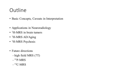 |
Things You Need to Know for In Vivo Molecular Imaging by MRS- perspectives from neuroradiology
Henry Mak
The first part of the talk gives a brief overview of the key basic concepts of proton MR spectroscopy such as chemical shift, J coupling and spectral editing, and some caveats in interpretation of MR spectroscopy findings. The second part of the talk emphasize its applications in brain tumors, neurodegenerative disease and psychosis. Finally, the future directions in MRS research are discussed.
|
|
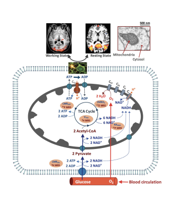 |
What Molecular Properties Can We Image using newly developed X-nuclear MRS methods?
Wei Chen, Xiao-Hong Zhu Video Permission Withheld
The advancement of ultrahigh-field (UHF) MRI technology (now reaching 10.5T for human scanner and beyond 16T for preclinical animal) has significantly improved imaging sensitivity, spectral and spatiotemporal resolutions. It accelerates new developments of in vivo MRS imaging technologies enabling quantitative and reliable assessment of various neurochemicals, metabolites, metabolic rates in healthy and diseased brain. This lecture will discuss newly developed X-nuclear MRS imaging methods for quantitatively imaging cerebral metabolic rates of glucose and oxygen, ATP production, TCA cycle and NAD redox ratio; and demonstrate promising applications for studying brain function and neuroenergetics under normal and diseased states at UHF.
|
|
| PET/MR
René Botnar
Cardiac PET/MR promises to combine multi-contrast and multi-parametric cardiac MRI that provides information on anatomy, left ventricular function, myocardial tissue viability, perfusion and oxygenation as well as fibrosis (T1), inflammation (T2) and iron (T2*) with the high sensitivity of PET for radiotracer detection, Thus, it promises to enable simultaneous assessment of molecular and cellular processes related to cardiovascular diseases such as atherosclerosis, post infarct remodelling, cardiomyopathy or heart failure. In this talk we will discuss both the promises but also the challenges related to cardiac PET/MR and show first results from clinical studies.
|
 Watch the Video