
Weekend Educational Session
Perfusion MRI
Session Topic: Perfusion MRI
Session Sub-Topic: Perfusion MRI
Weekend Course
ORGANIZERS: Jongho Lee, Fernando Calamante, Seung Hong Choi, Susan Francis
| Sunday Parallel 1 Live Q&A | Sunday, 9 August 2020, 14:00 - 14:30 UTC | Moderators: Henk Mutsaerts & Kathleen Schmainda |
Skill Level: Basic to Intermediate
Session Number: WE-12
Overview
This basic course will introduce the background on acquisition and analysis of arterial spin labeling (ASL), dynamic-susceptibility contrast (DSC) MRI, and dynamic contrast-enhanced (DCE) MRI. Furthermore, it will present some of their key clinical applications.
Target Audience
This course is designed for clinicians and scientists who are interested in imaging perfusion and permeability and have some knowledge about the potential utility of perfusion MRI in clinical applications.
Educational Objectives
As a result of attending this course, participants should be able to:
- Identify the most suitable acquisition strategies for measuring perfusion using ASL and DSC-MRI and for measuring permeability using contrast agents;
- Describe the key issues in the extraction of physiological parameters from ASL, DSC, and DCE data; and
- Describe how ASL principles can be used to measure physiological parameters beyond CBF.
| ASL: Basics
Maria A. Fernandez-Seara
The objective of this talk is to introduce the methodology of ASL data acquisition and analysis. Upon attendance, the audience should have a basic understanding of the technique and be able to choose the most adequate pulse sequence for a particular clinical application and the most appropriate acquisition parameters.
|
||
| ASL: Advanced Topics
Sophie Schmid
|
||
| Applications: ASL
Shalini Amukotuwa
|
||
| DSC-MRI: Basics
Amit Mehndiratta
Perfusion imaging using dynamic susceptibility contrast MRI is widely used in management of brain ischemia and stroke. It is based on change in T2* effect arising from local field inhomogeneity as the contrast flow from the tissue capillary network. Quantitative analysis is completely based on the mathematical understanding of the underlying capillary network model, either a simplified model based approach or a complex model free methods have been used in literature. There are pros and cons of each method, this lecture will discuss few of these important methods and there benefits and limitations.
|
||
| DSC-MRI in Neuroimaging: Sources of Errors & Artifacts
Atle Bjørnerud
|
||
 |
DCE-MRI: Acquisition
Jaeseok Park
This lecture is to provide fundamental principles of dynamics contrast enhanced (DCE) magnetic resonance imaging (MRI) from the perspective of acquisition. We will talk about T1-weighted dynamic data acquisition for DCE MRI, dynamic T1 (t) quantification with correction of B1 field inhomogeneities, and conversion of T1 maps to concentrations. We move on to delineation of contrast dynamics with time and discuss why we need to have high spatial and temporal resolution for accurate quantification of perfusion and microvascular permeability. Then, I will introduce some of the state-of-the-art, high resolution DCE MRI methods.
|
|
 |
DCE-MRI: Processing
Ka-Loh Li
This educational lecture discusses efforts in making accurate, high-spatial resolution, whole brain coverage microvascular parametric maps and reducing dosage of gadolinium based contrast agents (GBCA), using a newly developed dual-temporal resolution (DTR) DCE-MRI processing technique.
|
|
 |
Applications: Contrast-Based Perfusion MRI
Roh-Eul Yoo Video Permission Withheld
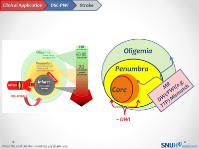
DSC-PWI is dependent on the susceptibility effect caused by paramagnetic gadolinium on T2*-weighted imaging. Dynamic contrast-enhanced (DCE) MR imaging is based on T1 shortening induced by a gadolinium-based contrast bolus passing through tissue. Various quantitative model-based and semiquantitative model-free pharmacokinetic parameters, that reflect microcirculatory structure and function, can be derived using the technique. This lecture will focus on the clinical applications of the contrast-based perfusion-weighted imaging techniques in neuroimaging (e.g. tumor, stroke, seizure).
|
 Watch the Video