SMRT Poster Presentations
Clinical
ISMRM & SMRT Annual Meeting • 15-20 May 2021

| SMRT Session | 03:00 - 04:00 | Moderators: |
S4.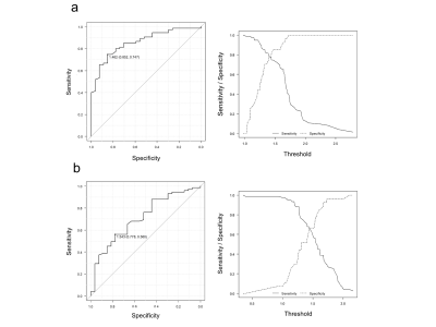 |
Investigation of new assessment method for the liver magnetic resonance imaging
Yasuo Takatsu1,2, Masafumi Nakamura3, Satoshi Kobayashi4, and Tosiaki Miyati4
1Department of Radiological Technology, Faculty of Health and Welfare, Tokushima Bunri University, Sanuki-city, Japan, 2Department of System Control Engineering, Graduate School of Engineering, Tokushima Bunri University, Sanuki-city, Japan, 3Department of Radiology, Otsu City Hospital, Otsu, Japan, 4Division of Health Sciences, Graduate School of Medical Sciences, Kanazawa University, Kanazawa, Japan
The hepatobiliary phase image using Gd–EOB–DTPA in the liver MRI is assessed by the quantitative liver–spleen contrast ratio (Q-LSC) , the cutoff value at which tumors can be easily determined is 1.5. However, Q-LSC is found unsuitable for cases of splenectomy and when there is splenic deposition of Gamna–Gandy bodies. Therefore, the quantitative liver-portal vein contrast ratio (Q-LPC) is useful instead of Q-LSC. The cutoff value of Q-LPC was at 1.462, the sensitivity and the specificity were higher than Q-LSC at the cutoff value. Q-LPC cutoff value can be used for hepatobiliary phase MR image evaluation.
|
||
S11.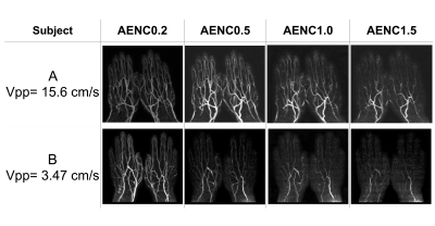 |
Non-gated and non-enhanced MR Angiography of the Hand using enhanced acceleration selective arterial spin labeling (eAccASL)
Misaki Saito1, Shuhei Shibukawa1, Natsuo Konta1, Takuya Hara2, Takakiyo Nomura2, Makoto Obara3, Isao Muro1, and Tetsu Niwa2
1radiological technology, Tokai university hospital, Isehara, Japan, 2Tokai University School of Medicine, Isehara, Japan, 3Philips Japan, Tokyo, Japan
This study was conducted to optimize the enhanced Acceleration-Selective Arterial Spin Labeling (eAccASL) for non-gated hand MRA. The MRA images obtained using four acceleration encoding (AENC) were assessed by visual evaluation for arterial depiction and venous contamination. The blood flow velocity obtained by phase-contrast cine was compared with the visual evaluation. It was found that the smaller the AENC (i.e. stronger motion sensitize gradients), the higher venous contamination and higher arterial visualization. The tendency of high visual evaluation was found in subjects with fast blood flow. The AENC of 0.5 or 1.0 was optimized for non-gated MRA of the hand.
|
||
S12.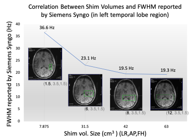 |
The Effect of Enlarging Shim Volume on B0 homogeneity of MRS Voxel on 7T
Huijun Liao1, Eduardo Jorge Uribe Coello1, Wufan Zhao1, Han Sam Jiang1, and Alexander Lin1
1Center for Clinical Spectroscopy, Brigham and Women's Hospital, Boston, MA, United States
B0 homogeneity is important for clinical spectroscopy but is highly region dependent and time consuming. Enlarging shim volume could be a quick and easy way to improve B0 homogeneity in the clinical practice of spectroscopy and the goal of this study is test if this method is beneficial. We used different methods to measure the potential improvement in B0 homogeneity in different brain regions. While our results showed that increasing shim volumes did not improve spectral linewidths or data quality, we demonstrate a robust method of measuring B0 homogeneity in clinical spectroscopy.
|
||
S25. |
Differential Diagnosis of uterine leiomyoma pathological subtypes: A Conditional Inference Tree Model Based on MRI-T2WI
Chao Wei1 and Jiangning Dong1
1Anhui provincial tumor hosptial, Hefei, China
The establishment and verification of conditional inference tree model based on based on the MRI-T2WI radiomics has high clinical value in the identification of three common pathological subtypes of uterine leiomyoma.
|
||
S30.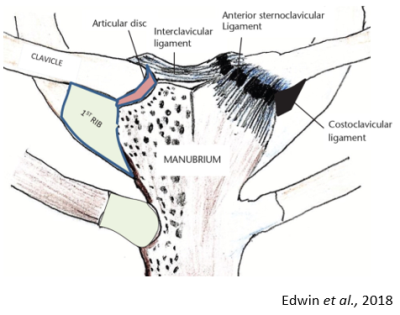 |
Depicting capsular injury and disc tears of the sternoclavicular joint with the use of direct MRI arthrography
Helen Harvey1
1MRI, Cambridge University Hospitals, Cambridge, United Kingdom Advances in technology in MRI have allowed for the improved spatial resolution of small joints like the sternoclavicular joint (SCJ). We have shown that through the use of arthrography and MRI we can provide better visualisation of SCJ anatomy, disc tears and other pathologies. Helping to increase knowledge and greatly improve treatment and surgical planning options for the patient. This presentation aims to understand the techniques for MRI arthrography of the SCJ, review anatomy demonstrated on SCJ arthrography and demonstrate common pathologies of the SCJ. |
||
S39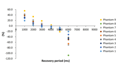 |
A Single Breath-Hold Acquisition of Three-Dimensional T1 Mapping Using Look Locker sequence for Assessing Crohn Disease: A Phantom Study Video Permission Withheld
Daisuke Morimoto-Ishikawa1,2, Tomoko Hyodo3, Shigeyoshi Saito2, Yu Ueda4, Masato Ohmi2, and Kazunari Ishii3
1Radiology Center, Kindai University Hospital, Osaka-Sayama, Japan, 2Department of Medical Physics and Engineering, Osaka University Graduate School of Medicine, Suita, Japan, 3Department of Radiology, Kindai University, Osaka-Sayama, Japan, 4Philips Japan, Tokyo, Japan
We investigated the single breath-hold three-dimensional (3D) Look-Locker (LL) sequence in nine different concentrations phantoms, which can be used to evaluate Crohn's disease (CD). Percentage errors between T1 relaxation time obtained by varying inversion recovery pulse interval and recovery period in the LL sequence and T1 relaxation time obtained by the Inversion Recovery sequence were calculated. With the combination of inversion recovery pulse interval of 7000ms and recovery period of 4000ms, the LL sequence can calculate T1 relaxation time within 10% percentage errors in the fastest imaging time of 21 seconds.
|
||
| S41. | Detection of Sacroiliac Joint Lesions in Axial Spondyloarthritis:Utility of Synthetic MRI
Ke Zhang1 and Guobin Hong2
1Radiology, The Fifth Affiliated Hospital,Sun Yat-sen University, Zhuhai, China, 2Radiology, the Fifth Affiliated Hospital, Sun Yat-sen University, Zhuhai, China
The current diagnosis of the sacroiliac joint lesions with axSpA is most concentrated on qualitative or semi-quantitative methods.There is an urgent need for a quantitative method that can objectively and accurately evaluate the severity of the disease.Synthetic MRI (MAGiC) can generate multiple contrast images and quantitative maps simultaneously based on the same scan.To our knowledge,this study is the first to investigate the potential practicality of synthetic MRI for assessing sacroiliac joint lesions of participants with axSpA.In this study we hypothesized that synthetic MRI could be used for the qualitative and quantitative diagnosis of sacroiliac joints lesions of participants with axSpA.
|
||
S74.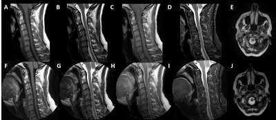 |
Optimization of Radial K-space Sampling Techniques for a Comprehensive Motion-free Cervical Spine MRI Protocol
Brian Johnson1, Jonathan Chia1, Dave Hitt1, Rob Lay1, Tom Lowe1, Michael Pawlak1, John Penatzer1, James Snicer1, Marcie Stopchinski1, Gregory Thomas1, Kristen Williams1, and Paul Worthington1
1Philips Healthcare, Gainesville, FL, United States
Consistent high-quality magnetic resonance imaging (MRI) of the cervical spine still remains challenging because of the inherent small anatomical structures and degradation of image quality due to motion artifacts1. Motion artifacts can arise from several sources including swallowing, respiration, cerebrospinal fluid (CSF) pulsation, blood flow, and bulk patient movement. MRI of the cervical spine is one the highest performed exams globally. Here we present a complete cervical spine MRI protocol utilizing 2D and 3D radial k-space sampling techniques which are inherently motion insensitive to increase image quality.
|
||
S78.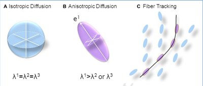 |
Diffusion: What is it and Why it is so Important in MR imaging
Gail H Kohls1, Robert Shih2, J Kevin Demarco2, and Maureen Hood2
1Radiology, USU/WRNMMC, MIDDLETOWN, MD, United States, 2Radiology, WRNMMC, Bethesda, MD, United States
The concepts behind diffusion imaging are complicated. As Technologist/Radiographers we use these finished pulse sequences in our daily work, but we do not always have the background information of how each sequence is developed and why it is an important part of the imaging we perform. This abstract is an attempt give the Technologist/Radiographer a better understanding of the key concepts of diffusion and how the diffusion sequences such as Diffusion Weighted Imaging (DWI) and Diffusion Tensor Imaging (DTI) measure water movement.
|
||
| S80. | A finding of Female Adnexal Tumour of probable Wolffian Origin (FATWO) on magnetic resonance imaging and histology - a rare neoplasm: Case report
Petronella Samuels1, Paul Scholtz2, Judith Whittaker3, and Sally Candy4
1Cape Universities Body Imaging Centre, University of Cape Town, Cape Town, South Africa, 2Department of Radiology, Morton & Partners Radiology, Cape Town, South Africa, 3Anatomical Pathology, Lancet Laboratories, Cape Town, South Africa, 4Division of Diagnostic Radiology, Groote Schuur Hospital and University of Cape Town, Cape Town, South Africa
We present the MRI and histological findings in a case of Female Adnexal Tumour of Wolffian Origin (FATWO) / WAT (Wolffian Adnexal Tumour), a rare adnexal neoplasm. This mass, located in the right broad ligament demonstrated diffusely low signal intensity on both T2WI and T1WI, peripheral restriction on DWI, peripheral enhancement and central hypo-enhancement post Gadolinium. An awareness of this condition and its MRI findings will ensure that appropriate immunohistochemical staining is performed. The presence of restriction may be helpful in predicting tumour behaviour, surgical approach and postoperative management. |
The International Society for Magnetic Resonance in Medicine is accredited by the Accreditation Council for Continuing Medical Education to provide continuing medical education for physicians.