SMRT Poster Presentations
Research
ISMRM & SMRT Annual Meeting • 15-20 May 2021

| SMRT Session | 03:00 - 04:00 | Moderators: |
S5. |
MRI Safety for Leave-on Powdered Hair Thickeners: Measurement of Deflection Force and MRI Artifact
Norio Hayashi1, Akio Ogura1, Atsuya Fuju2, Tomokazu Takeuchi3, Yusuke Sato4, Maiko Hashimoto5, and Masahiko Takahashi5
1Radiological Technology, Gunma Prefectural College of Health Sciences, Maebashi, Japan, 2Radiology, Kiryu Kosei General Hospital, Kiryu, Japan, 3Graduate School of Radiological Technology, Gunma Prefectural College of Health Sciences, Maebashi, Japan, 4Radiology, Gunma University Hospital, Maebashi, Japan, 5Isesaki Municipal Hospital, Isesaki, Japan
The purpose of this study was to evaluate the attraction force and image artifacts of 17 different leave-on powdered hair thickeners from 7 vendors. Displacement force was measured by the ASTM F2052, and MRI artifact was estimated by the ASTM F2052. There were some types of leave-on powdered hair thickener that do contain magnetic materials. They were highly susceptible to mechanical effects and will cause more image artifacts, so it must be removed before the examination.
|
||
S7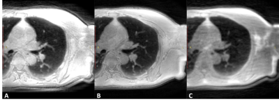 |
Optimized 3D ultrashort TE protocol for lung imaging Video Permission Withheld
CHIKARA NODA1, Chia Ying Liu2, Jason Ortman1, Bharath Venkatesh Ambale3, Webster Stayman4, Yoshimori Kassai5, and Joao A.C. Lima1
1Cardiology, Johns Hopkins University, Baltimore, MD, United States, 2Canon Medical Research, Cleveland, OH, United States, 3Radiology, Johns Hopkins University, Baltimore, MD, United States, 4Biomedical Engineering, Johns Hopkins University, Baltimore, MD, United States, 5Canon Medical Systems Corporation, Otawara, Japan
3D radial Ultrashort echo time sequence is a promising technique for lung imaging but the protocol has not been optimized. We examined the parameters including number of trajectories and flip angles using a lung phantom. In-vivo images were obtained and evaluated based on the parameters optimized in the phantom study. UTE acquisition in axial rather than coronal direction produced better quality of images. Breath holding acquisition may be an option in case patients cannot tolerate long scan time.
|
||
| S19. | Optimizing dark blood late gadolinium enhancement in cardiac magnetic resonance imaging.
Hannah Bergman1, Alaine Berry1, and Ben Statton1
1MRC, Imperial College, London, United Kingdom
The dark blood late gadolinium enhancement technique has shown improved delineation of sub-endocardial infarcts by nulling both the blood pool and the normal myocardium. Optimal image quality and consistency depends on a number of factors, including gadolinium dose and the delay time before imaging. In this study we sought to determine whether gadolinium dose and delay time before imaging, affected the nulling of the blood pool and myocardium.
|
||
S20.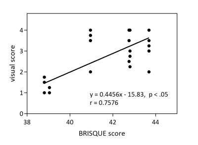 |
Quantitative evaluation of metal artifacts in 3-T MRI by using Blind/Referenceless Image Spatial Quality Evaluator (BRISQUE): a phantom study
Wakiko Tani1, Yutaka Katayama2, Tomoaki Yamakawa1, Shintaro Horii1, Ryuji Shimada1, Yuichiro Somiya1, and Akiko Kusaka1
1Center for Radiology and Radiation Oncology, Kobe University Hospital, Kobe, Japan, 2Division of Radiological Technology, Osaka City University Hospital, Osaka, Japan
No-reference image quality assessment (NR-IQA) model called Blind/Referenceless Image Spatial Quality Evaluator (BRISQUE) is one of the objective evaluations of images. To investigate whether BRISQUE can be used to quantify artifacts on MRI images, we compared the BRISQUE score with the visual score. The BRISQUE score was reduced and the visual score was improved by increasing bandwidths from 51 kHz to 307 kHz. Pearson correlation coefficient was 0.7576 and BRISQUE score was associated with the visual score (P < .05). This study suggests that BRISQUE can quantitatively evaluate metal artifacts.
|
||
S21.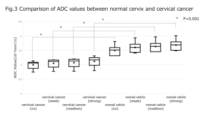 |
Compressed Sensing for Turbo Spin-echo Diffusion-weighted Imaging: Influence of Denoising Level on the Signal Intensity and Apparent Diffusion Coefficient Values in Cervical Cancer
Yuichiro Somiya1, Yoshiko Ueno2, Shintaro Horii1, Ryuji Shimada1, Keitaro Sofue2, Naoki Yoshida1, Wakiko Tani1, Akiko Kusaka1, and Takamichi Murakami2
1Center of Radiology and Radiation Oncology, kobe university hospital, kobe, Japan, 2Department of Radiology, Kobe University Graduate School of Medicine, kobe, Japan
This study evaluated the effect of denoising level on signal intensity (SI) and apparent diffusion coefficient (ADC) values of diffusion-weighted imaging using the single-shot turbo spin-echo sequence (TSE-DWI) with compressed sensing (CS) in cervical cancer. As the denoising level increased, the SI of cervical cancer tended to decrease, and the ADC values tended to increase on TSE-DWI with CS. Nevertheless, at each denoising level, the ADC values of cervical cancer were significantly lower than those of normal uterine cervix.
|
||
S31.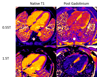 |
Comparison of cardiac T1 mapping on a high-performance 0.55T scanner and a conventional 1.5T scanner
Christine Mancini1, W. Patricia Bandettini1, Peter Kellman1, Hui Xue1, and Adrienne E. Campbell-Washburn1
1NHLBI, National Institutes of Health, Bethesda, MD, United States
This abstract is a comparison of native and post contrast T1 mapping and ECV of the myocardium using a conventional 1.5T scanner and a high-performance 0.55T scanner.
|
||
S34.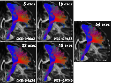 |
Effects of image quality deterioration and data shortage on automatic white matter bundle segmentation by diffusion magnetic resonance imaging
Yuichi Suzuki1, Tsuyoshi Ueyama1, Takahiro Iwasaki1, Jiro Sato1, Hideyuki Iwanaga1, and Osamu Abe1
1Department of Radiology, THe University of Tokyo Hospital, Tokyo, Japan
We investigated the effect of image quality deterioration and data shortage of DWI on automatic white matter bundle segmentation. We set the gold standard (GS) data without SMS and MPG = 64 axes. Two kinds of comparisons were made in comparison with GS data. The one was with/without the SMS, and the other eliminated some acquired data from GS. We analyze the data only from the first 8, 16, 32 and 48 axes of MPG in the GS. There was almost no difference between SMS of 2 and 3. For MPG, 16 axes provided results comparable to the GS.
|
||
S44.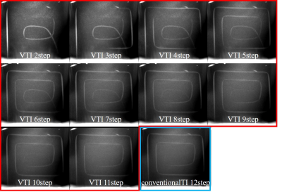 |
Optimization of Variable TI 4D ultrashort TE MR Angiography: A Numerical Simulation and Phantom Study
Toshiya Akatsu1, Haruyuki Fukuchi2,3,4, Kei Fukuzawa4, Nao Takano1, Yutaka Ikenouchi3, Michimasa Suzuki3, Kohji Kamagata3, Akihiko Wada3, Osamu Abe2, and Shigeki Aoki3
1Department of Radiology, Juntendo University Hospital, Tokyo, Japan, 2Department of Radiology, Graduate School of Medicine, University of Tokyo, Tokyo, Japan, 3Department of Radiology, Juntendo University Graduate School of Medicine, Tokyo, Japan, 4Department of Radiology, Toranomon Hospital, Tokyo, Japan
We implemented the new Variable TI method to multi-phase ASL 4D UTE-MRA in order to improve the visibility of hemodynamic flow. We performed a numerical simulation and a phantom study to find the optimized condition. It was found that a 50 % reduction of TI steps increases 50 % of signal from the flow volume without impairment of structural information.
|
||
S59.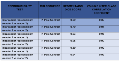 |
Assessing the reproducibility of semi-automated segmentation methods on post-operative T1 post contrast and FLAIR MRI of GBM.
Olga V Fadeeva Da Costa1,2, Shah Islam2,3, Mark M Boubnovski3, Eric Aboagye3, and Adam D Waldman4,5
1Department of Surgery & Cancer, Cancer Imaging Centre, Hammersmith Campus, Imperial College, London, United Kingdom, 2Imaging Department, MRI Unit, Hammersmith Hospital, Imperial College Healthcare NHS Trust, London, United Kingdom, 3Department Of Surgery & Cancer, Cancer Imaging Centre, Hammersmith Campus, Imperial College, London, United Kingdom, 4Department of Brain Sciences, Hammersmith Campus, Imperial College, London, United Kingdom, 5Centre for Clinical Brain Sciences, University of Edinburgh, Edinburgh, United Kingdom
Glioblastoma (GBM) account for ~70% of adult primary brain tumours. Interrogation of post-operative disease residuum and peritumoral microenvironment is necessary for imaging biomarker development. This involves accurate delineation and segmentation of these particular regions of interest. A total of 35 post-operative T1 post contrast and T2 FLAIR MRI’s underwent segmentation by two independent readers. There was poor inter reader reproducibility in segmentations of disease residuum on post contrast T1 images, although this improved was higher for on segmentation of abnormal regions on FLAIR. Strong intra reader reproducibility suggests systematic error effects in identifying regions of interest.
|
||
S60.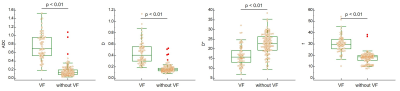 |
Osteoporotic vertebral fracture analysis with intravoxel incoherent motion: a preliminary study
Hiroyuki Takashima1,2, Rui Imamura1, Tsuneo Takebayashi3, Yasuhisa Abe3, Izaya Ogon2, Hiroshi Oguma3, Yoshihiro Akatsuka1, and Toshihiko Yamashita2
1Division of Radiology and Nuclear Medicine, Sapporo Medical University Hospital, Sapporo, Japan, 2Department of Orthopaedic Surgery, Sapporo Medical University School of Medicine, Sapporo, Japan, 3Department of Orthopaedic Surgery, Sapporo Maruyama Orthopedic Hospital, Sapporo, Japan
The parameters of vertebral fracture (VF) and without fracture using intravoxel incoherent motion (IVIM) were analyzed. In addition, the usefulness of using IVIM for VF was investigated. The subjects of this study were 52 patients with VF in the acute phase. As a result, the ADC, D, and f of VF were significantly higher than without fracture. Meanwhile, the D* of VF were significantly lower than without fracture. In the future, new findings may be obtained by analyzing the IVIM parameters and VF prognosis.
|
||
S81.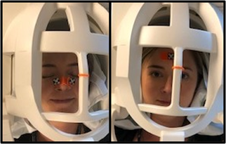 |
Optimizing a Motion Tracking Marker for Pediatric Patients
Kristina Mary Pelkola1,2, Onur Afacan1,2, Tess E. Wallace1,2, Pauline Connaughton1, Jenna McKay1, Joseph Zmuda1, Camilo Jaimes1, and Simon K. Warfield1,2
1Radiology, Boston Children's Hospital, Boston, MA, United States, 2Computational Radiology Laboratory, Boston Children's Hospital, Boston, MA, United States
Magnetic Resonance Imaging (MRI) can be challenging for pediatric patients due to the large tunnel and loud noises. Anxiety and fear can be sparked causing them to be uncooperative and unable to hold still [1,2]. These exams are often plagued with motion artifacts resulting in poor diagnostic image quality and a challenge for Radiologists to interpret [3]. The administration of sedation or anesthesia is costly, potentially harmful, and doesn't eliminate motion artifacts due to breathing and uncontrollable muscle spasms [4]. There is an unmet need for alternative methods to enable diagnostic imaging exams in the presence of motion.
|
||
S90.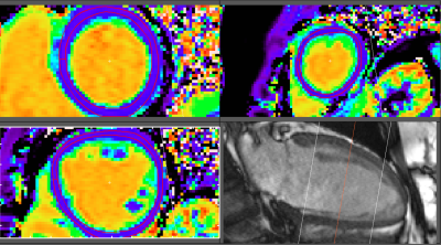 |
Deep Inspiration Breath-Hold Radiotherapy does not induce MRI detectable LV functional and structural changes in patients with left-sided breast cancer – A six months follow-up
Xin Dong1,2, Arnold Ng3,4, Sharon Watson5, Harish Sharma5, Graham Galloway1,2,6, and Margot Lehman5
1Queensland University of Technology, Brisbane, Australia, 2Translational Research Institute, Woolloongabba, Australia, 3Cardiology, Princess Alexandra Hospital, Brisbane, Australia, 4University of New South Wales, Sydney, Australia, 5Radiation Oncology, Princess Alexandra Hospital, Brisbane, Australia, 6The University of Queensland, St Lucia, Australia
The usefulness of T1 and ECV mappings in the context of radiation cardiotoxicity has yet to be studied. In the current study, we conducted a longitudinal CMR study on 30 females with left-sided breast cancer before and after Deep Inspiration Breast Hold (DIBH) radiotherapy (RT). The results showed patients who were treated with DIBH RT had no CMR detectable functional or structural myocardial changes 6 months following RT treatment. This has important implications on the long-term cardiac health of the growing number of women who are surviving or living with breast cancer.
|
||
S92.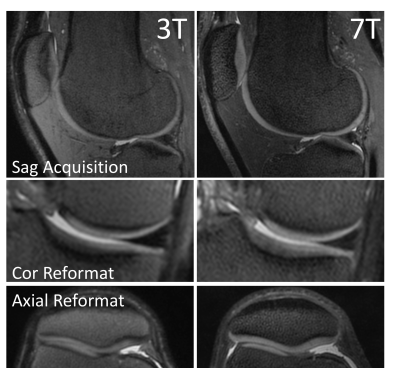 |
7T Knee MRI: Clinical Benefits and Challenges
Kevin Epperson1, Karla Epperson1, Garry Gold2, and Feliks Kogan3
1Radiology School of Medicine, Stanford University, Stanford, CA, United States, 2Radiology, Stanford Univeristy, Stanford, CA, United States, 3Radiology - SOM, Stanford Univeristy, Stanford, CA, United States
Diagnostic MRI scanning of the knee at 3T offers multiple contrasts and definition of anatomical structures, presenting pathology, and allows quantitative performance methods. MRI scanning at 7T of the knee presents challenges, however the ultra high field system offers improved resolution and SNR for visualization of small morphological changes and translation of quantitative methods when these challenges are satisfied.
|
The International Society for Magnetic Resonance in Medicine is accredited by the Accreditation Council for Continuing Medical Education to provide continuing medical education for physicians.