Combined Educational & Scientific Session
Imaging of Heart Failure
ISMRM & SMRT Annual Meeting • 15-20 May 2021

Combined Educational & Scientific Session
Imaging of Heart Failure
| Concurrent 3 | 12:00 - 14:00 | Moderators: Bruno Quesson & Lixian Zou |
| What Is Heart Failure?: Overview of Pathophysiology & Epidemiology
Daniel Messroghli
|
||
| Key Clinical Trials in Heart Failure
Matthew Lee
CMR has roles in the diagnosis and risk stratification of heart failure. CMR is the reference "gold" standard method of assessing cardiac structure and function. CMR reduces the sample size required in heart failure clinical trials. CMR has been used in several key heart failure trials to assess cardiac remodelling.
|
||
 |
The Role of MRI in Diagnosis & Management of Heart Failure
Kim-Lien Nguyen
Heart failure is a complex clinical syndrome with many causes. Although imperfect, the left ventricular ejection fraction (LVEF) serves as a surrogate marker for combined LV function and structural phenotyping of heart failure. Cardiac imaging provides information about structure, tissue composition, and function. As a diagnostic modality with high spatial resolution and no ionizing radiation exposure, cardiovascular MRI is valuable for accurate EF quantification and phenotypic characterization. However, several barriers exist for widespread implementation of quantitative MRI in heart failure. Examples of challenges include accessibility to and standardization of cardiovascular imaging pulse sequences for probing myocardial tissue structure and function.
|
|
 |
How Can MR Imaging Address Knowledge Gaps in Heart Failure?
Sebastian Kozerke
Cardiac Magnetic Resonance offers a multitude of contrast mechanisms across spatiotemporal scales to further improve diagnosis, stratification and interventions in heart failure patients. In this presentation the focus is put on hyperpolarized metabolic and diffusion tensor imaging to unravel the interplay of energy supply and myofiber contractile reserve in conjunction with biophysical modeling of left-ventricular function and dysfunction. It is shown that statistical learning and, in particular, physics-informed neural networks offer approaches for rapid personalization, detection and prediction of failing hearts.
|
| Concurrent 3 | 12:00 - 14:00 | Moderators: |
 |
0021.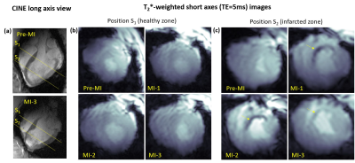 |
Visualization of Post-Infarction Cardiac Tissue Remodeling at 7T using T2* contrast: Longitudinal Study in a Pig Myocardial Infarction Model
Maxim Terekhov1, David Lohr1, Michael Hock1, Maya Bille1, Steffen Baltes1, Ibrahim A. Elabyad1, Florian Schnitter2, Julia Aures1, Theresa Reiter2, Wolfgang Bauer1,2, Ulrich Hofmann2, and Laura M. Schreiber1
1Chair of Molecular and Cellular Imaging, University Hospital Würzburg, Comprehensive Heart Failure Center, Wuerzburg, Germany, 2Department of Internal Medicine I, Cardiology, University Hospital Würzburg, Wuerzburg, Germany
Cardiac MRI at 7T is a developing methodology capable to increase physical sensitivity and spatial resolution of the cardiac MR-images. T2*-quantification at 7T is of particular interest because it is highly sensitive to cardiac microstructure and has the potential to improve data quality on cardiac tissue remodeling in both acute and chronic cardiac diseases compared to clinical B0-field. In this work, we present the results of a pilot study correlating the multi-slice T2*-data with late-gadolinium-enhancement and first-pass-perfusion results in the large animal model of myocardial infarction. 8 pigs measured prior and at three dates after inducing myocardial infarction are analyzed.
|
|
0022.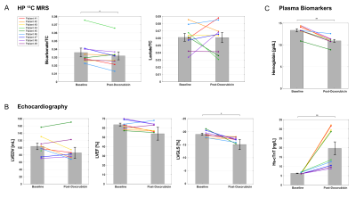 |
Effect of doxorubicin treatment on myocardial metabolism in patients with breast cancer
Jae Mo Park1,2,3, Galen D Reed4, Jeff Liticker1, William C Putnam5, Alvin Chandra6,7, Katarina Yaros6, Aneela Afzal1,6, James MacNamara6, Ronald G Hall5, Crystal E Harrison1, Alagar Muthukumar6, Colby Ayers6, James de Lemos6,
Craig R Malloy1,6, Hsiao-Ching Li6,7, Barbara Haley6,7, and Vlad G Zaha1,6
1Advanced Imaging Research Center, University of Texas Southwestern Medical Center, Dallas, TX, United States, 2Radiology, University of Texas Southwestern Medical Center, Dallas, TX, United States, 3Electrical and Computer Engineering, University of Texas at Dallas, Richardson, TX, United States, 4GE Healthcare, Dallas, TX, United States, 5Pharmacy Practice, Texas Tech University, Dallas, TX, United States, 6Internal Medicine, University of Texas Southwestern Medical Center, Dallas, TX, United States, 7Harold C. Simmons Comprehensive Cancer Center, University of Texas Southwestern Medical Center, Dallas, TX, United States
This study evaluates the feasibility of hyperpolarized 13C MRS for detection of early metabolic response in the myocardium of patients after anthracycline treatment. Patients with breast cancer were studied using hyperpolarized [1-13C]pyruvate before and after doxorubicin. Appearance of [13C]bicarbonate was significantly decreased after doxorubicin compared to the baseline exam while no change in [1-13C]lactate was detected. The reproducibility of hyperpolarized exams was examined by two injections of hyperpolarized [1-13C]pyruvate. The study demonstrates that hyperpolarized 13C spectra of the heart are sensitive to early injury of the mitochondria after doxorubicin therapy in patients with breast cancer in a reproducible manner.
|
||
0023.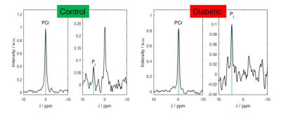 |
Increased Cardiac Pi in the Diabetic Heart Observed Using STEAM 31P MRS at 7T
Ladislav Valkovic1,2, Andrew Apps1, Jane Ellis1, Damian J Tyler1,3, Stefan Neubauer1, Albrecht Ingo Schmid4, Oliver J Rider1, and Christopher T Rodgers1,5
1Oxford Centre for Clinical Magnetic Resonance Research (OCMR), RDM Cardiovascular Medicine, University of Oxford, Oxford, United Kingdom, 2Department of Imaging Methods, Institute of Measurement Science, Slovak Academy of Sciences, Bratislava, Slovakia, 3Department of Physiology, Anatomy and Genetics, University of Oxford, Oxford, United Kingdom, 4High Field MR Center, Center for Medical Physics and Biomedical Engineering, Medical University of Vienna, Vienna, Austria, 5Wolfson Brain Imaging Centre, Department of Clinical Neurosciences, University of Cambridge, Cambridge, United Kingdom
Impaired cardiac energetics are characterized by a reduced phosphocreatine to adenosine-triphosphate ratio (PCr/ATP), however, changes in inorganic phosphate (Pi) may impact the Gibbs energy of ATP hydrolysis earlier in the disease process. Quantifying this in the diabetic heart may help explain latent diastolic dysfunction. Therefore, we used STEAM 31P-MRS at 7T to measure Pi/PCr in a type 2 diabetic (T2DM) cohort, and demonstrated an increased Pi/PCr in the diabetic human heart in comparison to healthy subjects. No correlation between PCr/ATP and Pi/PCr hints that multiple mechanisms contribute to these perturbations with candidates including impairment of CK flux and substrate inflexibility.
|
||
0024.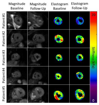 |
Left Ventricular Myocardial Stiffness Decreases after Stem Cell Therapy in Amyloidosis Patients: Monitored with Cardiac MR Elastography
Arvin Arani1, Jessica Magnuson1, Joshua D. Trzasko1, Yi Sui1, Kevin J. Glaser1, Armando Manduca1, Richard L. Ehman1, Sudhakar K. Venkatesh1, and Philip A. Araoz1
1Mayo Clinic, Rochester, MN, United States
Light chain (AL) cardiac amyloidosis is a disease where abnormal proteins are deposited in the heart tissue, commonly resulting in elevated myocardial stiffness. In cases with poor prognosis, autologous hematopoietic stem cell transplantation is used as a therapy. However, organ response monitoring is currently limited and challenging. The goal of this study is to evaluate the feasibility of using cardiac magnetic resonance elastography (MRE) to monitor changes in left ventricular (LV) myocardial stiffness after stem cell transplantation for treatment of AL amyloidosis. MRE detected a statistically significant (p=0.007) decrease in LV stiffness (mean decrease: 1.6±0.7 kPa, 17.0±7.4%) post therapy.
|
||
|
0025. |
Feasibility of single breath-hold CINE with combined Simultaneous Multi-Slice (SMS) and Region-Optimized Virtual (ROVir) coils
Daeun Kim1, Rodrigo A. Lobos1, Jaume Coll-Font2,3,4, Maaike van den Boomen2,3,4, John Conklin2,5,6, Jianing Pang7, Daniel Staeb8, Peter Speier9, Xiaoming Bi10, Brian Ghoshhajra5,6, Justin P. Haldar1, and Christopher T. Nguyen2,3,4
1Department of Electrical and Computer Engineering, University of Southern California, Los Angeles, CA, United States, 2A. A. Martinos Center for Biomedical Imaging, Department of Radiology, Massachusetts General Hospital, Charlestown, MA, United States, 3Cardiovascular Research Center, Cardiology Division, Massachusetts General Hospital, Charlestown, MA, United States, 4Department of Medicine, Harvard Medical School, Boston, MA, United States, 5Department of Radiology, Massachusetts General Hospital, Charlestown, MA, United States, 6Department of Radiology, Harvard Medical School, Boston, MA, United States, 7Siemens Medical Solutions USA Inc., Chicago, IL, United States, 8Siemens Healthcare Pty Ltd, Melbourne, Australia, 9Siemens Healthcare GmbH, Erlangen, Germany, 10Siemens Medical Solutions USA Inc., Los Angeles, CA, United States
Conventional clinical cardiac MRI protocols use a large number (>20) of breath-holds for capturing cinemagraphic (CINE) scans of the heart in various views. We hypothesize simultaneous multi-slice (SMS) CINE can be further accelerated using a reduced FOV and a novel approach based on Region-Optimized Virtual (ROVir) coils, which can potentially achieve single breath-hold whole heart CINE. We demonstrated the feasibility of combining SMS and ROVir for highly accelerated CINE imaging (8-fold reduced scan time), enabling single breath-hold whole ventricular acquisition. Single breath-hold SMS+ROVir whole-heart CINE yielded cardiac function parameters with no significant bias when compared to SMS CINE.
|
|
|
0026. |
Low Rank Motion Correction for free breathing first pass myocardial perfusion
Gastao Cruz1, Alina Hua1, Camila Munoz1, Tevfik Ismail1, Amedeo Chiribiri1, René M. Botnar1, and Claudia Prieto1
1School of Biomedical Engineering and Imaging Sciences, King's College London, London, United Kingdom
Conventional myocardial perfusion imaging requires free breathing acquisitions, with high spatial and temporal resolution. Leveraging the underlying redundant anatomic information to improve this multi-contrast application is challenging due to motion. Here a novel Low Rank Motion Corrected (LRMC) reconstruction is proposed to enable highly accelerated, motion corrected free breathing first pass myocardial perfusion imaging. This approach combines low rank subspace modelling (to resolve contrast) and non-rigid motion fields (to correct respiratory motion). The proposed approach successfully corrects respiratory motion and considerably improves image quality compared to conventional iterative SENSE reconstruction.
|
The International Society for Magnetic Resonance in Medicine is accredited by the Accreditation Council for Continuing Medical Education to provide continuing medical education for physicians.
 Watch the Video