Combined Educational & Scientific Session
Ebb & Flow: Perfusion & Permeability from Head to Toe
ISMRM & SMRT Annual Meeting • 15-20 May 2021

Combined Educational & Scientific Session
Ebb & Flow: Perfusion & Permeability from Head to Toe
| Concurrent 6 | 12:00 - 14:00 | Moderators: Mami Iima & Ananth Madhuranthakam |
 |
MR Perfusion in Glioma Management Video Permission Withheld
Kentaro Akazawa
Magnetic resonance (MR) perfusion identifies tumor angiogenesis or the proliferation of abnormal vessels in tumors. Of the various techniques devised for evaluating cerebral perfusion imaging, the dynamic susceptibility contrast (DSC) method has been employed most widely in clinical practice. DSC perfusion is used to generate hemodynamic parameters such as relative cerebral blood volume (rCBV). CBF maps can be used to assess neovascularity in tumors, which is thought to correlate with tumor grade and malignant histology. This talk will provide knowledge on how DSC perfusion can be useful in the glioma management in a variety of clinical conditions.
|
|
| Review of Perfusion & MR Placental Imaging
Oliver Wieben
|
||
| DCE-MR
Sadhna Verma
|
||
 |
Cardiac MR Perfusion: Updates
Frederick Epstein
First-pass MRI utilizes ECG-gated saturation-recovery gradient-echo or SSFP imaging applied immediately upon intravenous injection of gadolinium, enabling the visual or quantitative assessment of myocardial perfusion. For multislice coverage with high spatiotemporal resolution, acceleration is required. While parallel imaging is standard, compressed sensing, multiband, and/or non-Cartesian trajectories provide improvements, and deep learning may facilitate rapid reconstruction. Perfusion quantification is important in three-vessel and microvascular disease, and recent deep-learning-based pipelines promise to make quantification routine. Clinically, while superiority compared to SPECT was demonstrated in 2012, recently noninferiority compared to fractional flow reserve for guiding coronary revascularization was shown, representing another major advance.
|
| Concurrent 6 | 12:00 - 14:00 | Moderators: |
 |
0047.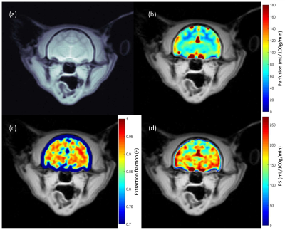 |
Measurement of Blood-Brain Barrier Disruption in Cats with an Inherited Neurodevelopmental Abnormality using Magnetization Transfer-ASL at 7T
Sultan Zaman Mahmud1,2, Emily C. Graff3,4, Douglas R. Martin4,5, Thomas S. Denney1,2, and Adil Bashir1,2
1Department of Electrical and Computer Engineering, Auburn University, Auburn, AL, United States, 2Auburn University MRI Research Center, Auburn University, Auburn, AL, United States, 3Department of Pathobiology, Auburn University, Auburn, AL, United States, 4Scott-Ritchey Research Center, Auburn University, Auburn, AL, United States, 5Department of Anatomy, Physiology and Pharmacology, Auburn University, Auburn, AL, United States
The blood-brain barrier (BBB) plays a vital role in regulating nutrient transport and acts as a barrier to potentially harmful molecules. Disruption of the BBB alters normal neurodevelopment and neuronal function. Protein enriched in astrocytes 15-kDa (PEA15) is crucial in normal neurodevelopment of cats, and cats with a PEA15 loss-of-function (PEA15-/-) have structural brain abnormalities and behavioral defects. We have previously demonstrated a non-invasive MRI technique to measure BBB permeability in humans. The goal of this study is to investigate if this technique can detect differences in microvascular cerebral blood flow and BBB in PEA15-/- cats compared to PEA15+/+.
|
|
|
0048.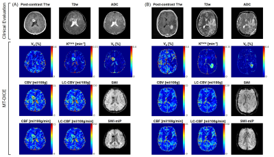 |
MR Multitasking-based Dynamic Imaging for Cerebrovascular Evaluation (MT-DICE): Further Development and Feasibility Study on Brain Cancer
Zhehao Hu1,2, Anthony Christodoulou1,2, Nan Wang1, Yibin Xie1, Tianle Cao1,2, Marcel Maya3, Wensha Yang4, Debiao Li1,2, and Zhaoyang Fan1,4,5
1Biomedical Imaging Research Institute, Cedars-Sinai Medical Center, Los Angeles, CA, United States, 2Department of Bioengineering, University of California, Los Angeles, Los Angeles, CA, United States, 3Department of Imaging, Cedars-Sinai Medical Center, Los Angeles, CA, United States, 4Department of Radiation Oncology, Keck School of Medicine, University of Southern California, Los Angeles, CA, United States, 5Department of Radiology, Keck School of Medicine, University of Southern California, Los Angeles, CA, United States
DSC-MRI and DCE-MRI provide perfusion- and permeability-related parameters, respectively, and are evolving as increasingly common modalities for evaluating a variety of brain cancer diseases. Their different but complementary information may form a more complete basis for evaluation of the complex and heterogeneous tumor microenvironment. However, acquiring both in one exam requires two separate scans as well as two contrast injections. In this work, we propose an MR MultiTasking based Dynamic Imaging for Cerebrovascular Evaluation (MT-DICE) technique that provides DCE- and leakage-corrected DSC-MRI parameters simultaneously with one 7.6-minute scan and a single-dose contrast injection.
|
|
|
0049.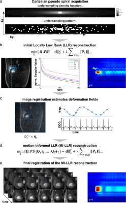 |
Free-Breathing Motion-Informed Quantitative 3D Myocardial Perfusion Imaging
Tobias Hoh1, Valery Vishnevskiy1, Maximilian Fuetterer1, and Sebastian Kozerke1
1Institute for Biomedical Engineering (IBT), University and ETH Zurich, Zurich, Switzerland
Three-dimensional perfusion CMR requires acceleration methods to enable whole-heart coverage in short acquisition windows, which often rely on data correlation among adjacent time-frames. However, in free-breathing examinations, respiratory motion leads to inconsistencies in the shared data and compromises image quality. In this work, non-rigid organ motion is incorporated into a patch-based locally low-rank reconstruction algorithm as a transformation displacement field for each time frame. This motion-informed locally low-rank reconstruction, combined with Cartesian pseudo-spiral k-t undersampling, is proposed as a dual-sequence acquisition framework to enable quantitative free-breathing whole-heart perfusion CMR. Feasibility is demonstrated in simulations, and volunteers in rest and stress.
|
|
 |
0050.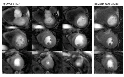 |
Whole heart SMS-bSSFP perfusion imaging with high resolution at 1.5T
Sarah McElroy1, Giulio Ferrazzi2, Muhummad Sohaib Nazir1, Carl Evans1, Filippo Bosio1, Nabila Mughal1, Karl P Kunze3, Radhouene Neji3, Peter Speier4, Daniel Stäb5, Christoph Forman4, Pier Giorgio Masci1, Reza Razavi1, Amedeo
Chiribiri1, and Sébastien Roujol1
1King's College London, London, United Kingdom, 2IRCCS San Camillo Hospital, Venice, Italy, 3Siemens Healthcare Limited, Frimley, United Kingdom, 4Siemens Healthcare GmbH, Erlangen, Germany, 5Siemens Healthcare Ltd, Melbourne, Australia
First-pass cardiac magnetic resonance (CMR) perfusion imaging is widely used for non-invasive assessment of coronary artery disease (CAD). Conventional CMR perfusion sequences are limited in spatial coverage and resolution. We have developed a SMS-bSSFP sequence with a total acceleration factor of 16 (multiband (MB) acceleration of 3 x in-plane acceleration of 5.5) and compressed sensing reconstruction to enable 1.5 T CMR perfusion with whole-heart coverage and high (1.4 x 1.4 mm2) in-plane spatial resolution. The results from a preliminary study in 6 patients comparing the proposed acquisition against a conventional 3-slice acquisition are presented.
|
|
0051.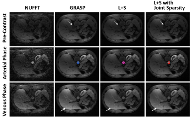 |
Improved Dynamic Contrast Enhanced MRI Using Low Rank with Joint Sparsity Reconstruction
Jichang Zhang1, Faisal Najeeb2, Xinpei Wang1, Pengfei Xu1, Hammad Omer2, Penny Gowland 3, Sue Francis3, Paul Glover3, Richard Bowtell3, and Chengbo Wang1
1SPMIC, The University of Nottingham Ningbo China, Ningbo, China, 2COMSATS University Islamabad, Islamabad, Pakistan, 3SPMIC, The University of Nottingham, Nottingham, United Kingdom
This work presents a free breathing Dynamic Contrast Enhanced MRI (DCE-MRI) reconstruction method called L+S (Low rank plus sparse) with joint sparsity, which improved dynamic contrast performance through integrating an additional temporal Fast Fourier Transform (FFT) constraint by extending the standard L+S decomposition method. Fast Composite Splitting Algorithm (FCSA) is implemented to solve the L+S optimization problem in proposed method, and to minimize the computation complexity from joint sparsity constraints. The proposed method achieved high spatial-temporal resolution, high reconstruction efficiency and improved dynamic contrast simultaneously when comparing with other methods in reconstructing a simulated phantom dataset and a DCE-MRI dataset.
|
||
0052.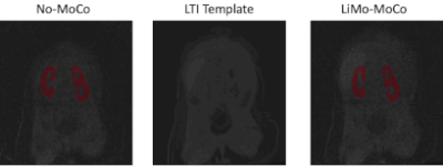 |
Linear Time Invariant Model based Motion Compensation for Renal Function Estimation with DCE-MRI
Jaume Coll-Font1, Onur Afacan1, Jeanne Chow1, Richard S Lee1, Simon K Warfield1, and Sila Kurugol1
1Boston Children's Hospital and Harvard Medical School, Boston, MA, United States
Heavy breathing or large bulk motion of infants during acquisition of renal DCE-MRI causes misalignment between volumes, degrades image quality, and reduces the accuracy of estimated quantitative parameters of kidney function. We proposed a robust LTI model-based registration algorithm for motion correction, which improved the temporal stability of the concentration time curves, resulting in improved estimation of tracer kinetic model parameters such as renal filtration rate.
|
The International Society for Magnetic Resonance in Medicine is accredited by the Accreditation Council for Continuing Medical Education to provide continuing medical education for physicians.
 Watch the Video