Combined Educational & Scientific Session
Breast MRI as a Problem-Solving Tool
ISMRM & SMRT Annual Meeting • 15-20 May 2021

Combined Educational & Scientific Session
Breast MRI as a Problem-Solving Tool
| Concurrent 3 | 16:00 - 18:00 | Moderators: Linda Moy & Naoko Mori |
| When to Use Advanced Sequences & AI
Katja Pinker-Domenig
This presentation will explain the concept and methodology of AI-enhanced multiparametric MRI, summarize the current applications in breast cancer and address its challenges and limitations.
|
||
| Difficult Cases the Breast Radiologist Should Know Video Permission Withheld
Silvia Perez
|
||
 |
Approach to High-Risk Screening
Eun Sook Ko
High-risk women can be identified by genetic testing, mathematical risk prediction models and risk factor assessment. Reasons supporting the use of MRI for breast cancer screening may be summarized in two ways: the performance of MRI and the biologic characteristics of MRI-detected cancer. In general, annual screening is recommended by the ACS for BRCA mutation, any untested first degree relative of a known mutation carrier, anyone with a > 20% lifetime risk of developing breast cancer, history of thoracic radiation, or a known syndrome (Li-Fraumeni or Cowden’s). Abbreviated MRI, ultrafast MRI, or DWI could be additional option for high-risk screening.
|
|
| How to Evaluate Treatment Response
Nola Hylton
|
| Concurrent 3 | 16:00 - 18:00 | Moderators: |
0139.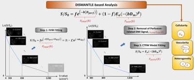 |
A Hybrid DWI Approach for Simultaneous Assessment of Cellularity, Vascularity, and Heterogeneity of Breast Lesions
Muge Karaman1,2, Yangyang Bu3,4, Guangyu Dan1,2, Zheng Zhong1,2, Qingfei Luo1, Shiwei Wang3,4, Changyu Zhou3,4, Weihong Hu3,4, X. Joe Zhou1,2,5, and Maosheng Xu3,4
1Center for Magnetic Resonance Research, University of Illinois at Chicago, Chicago, IL, United States, 2Department of Bioengineering, University of Illinois at Chicago, Chicago, IL, United States, 3The First Clinical Medical College, Zhejiang Chinese Medical University, Hangzhou, China, 4Department of Radiology, The First Affiliated Hospital of Zhejiang Chinese Medical University, Hangzhou, China, 5Departments of Radiology and Neurosurgery, University of Illinois at Chicago, Chicago, IL, United States
Breast cancer is the second most common cancer among women. Breast tissue has a variety of structural features leading to different tissue properties which are collectively linked to the pathologic state of the breast lesions. In this study we demonstrate a hybrid DWI approach for simultaneous assessment of tissue cellularity, vascularity, and heterogeneity (DISMANTLE) based on intravoxel incoherent motion (IVIM) and continuous-time random walk (CTRW) diffusion models. Our results have shown that DISMANTLE improved the differentiation among benign and malignant breast lesions compared to the classical implementation of either IVIM or CTRW model.
|
||
0140.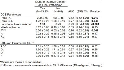 |
Quantitative DCE and DW-MRI to Evaluate Suspicious Mammographic Calcifications: Results from a Single Institution Prospective Clinical Trial
Janis M. Yee1, Daniel S. Hippe1, Michael Hirano1, Bonny Chau1, Debosmita Biswas1, Anum S. Kazerouni1, Mary Lynn Bryant1, Isabella Li1, Jennifer Xiao1, Wei Huang2, Savannah C. Partridge1, and Habib Rahbar1
1Radiology, University of Washington, Seattle, WA, United States, 2Oregon Health & Science University, Portland, OR, United States
Mammographic calcifications remain a diagnostic dilemma with low positive predictive value for malignancy. Our study investigated the use of quantitative DCE and DW-MRI metrics to problem solve suspicious calcifications prior to biopsy. In patients with a suspicious enhancing MRI correlate, malignant lesions exhibited higher peak PE, peak SER, functional tumor volume, and Ktrans. Basic and advanced 3D DW-MRI parameters did not yield statistically significant discriminatory values in this small pilot study. Use of quantitative MRI features shows potential to reduce the number of unnecessary biopsies for suspicious mammographic calcifications among the subset that demonstrate suspicious enhancement.
|
||
 |
0141.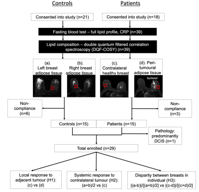 |
Deregulation of lipid composition in peri-tumoural adipose tissue in postmenopausal patients with breast cancer
Sai Man Cheung1, Vasiliki Mallikourti1, Tanja Gagliardi2,3, Ehab Husain4,5, Yazan Masannat5,6, Steven D Heys6, and Jiabao He1
1Institute of Medical Sciences, University of Aberdeen, Aberdeen, United Kingdom, 2Clinical Radiology, Aberdeen Royal Infirmary, Aberdeen, United Kingdom, 3Radiology, Royal Marsden Hospital, London, United Kingdom, 4Pathology Department, Aberdeen Royal Infirmary, Aberdeen, United Kingdom, 5School of Medicine, University of Aberdeen, Aberdeen, United Kingdom, 6Breast Unit, Aberdeen Royal Infirmary, Aberdeen, United Kingdom
Deregulation of lipid metabolism has been shown in BRCA1/2 genetic mutation carriers. Mammary adipose tissues in postmenopausal women are the primary sites of oestrogen production linked to tumour initiation and progression. Therefore, lipid composition in postmenopausal breast plays a key role in breast cancer monitoring and subsequent development of prevention strategies. Previous studies focused on cell or animal models and invasive lipid extraction methods, while conventional MRS is inadequate in complete lipid composition measurement. We hypothesised that lipid composition in peri-tumoural breast adipose tissue is affected by the presence of tumour in postmenopausal women, using a non-invasive 2D MRS approach.
|
|
|
0142.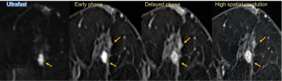 |
Ultrafast DCE MRI for post-NST evaluation of breast cancer
Maya Honda1, Masako Kataoka1, Rie Ota1, Mami Iima1,2, Akane Ohashi3, Kanae Kawai Miyake1, Marcel Dominik Nickel4, Yosuke Yamada5, Masakazu Toi6, and Yuji Nakamoto1
1Graduate School of Medicine, Kyoto University, Kyoto, Japan, 2Institute for Advancement of Clinical and Translational Science (iACT), Kyoto University Hospital, Kyoto, Japan, 3Department of Radiology, National Hospital Organization Kyoto Medical Center, Kyoto, Japan, 4MR Application Predevelopment, Siemens Healthcare GmbH, Erlangen, Germany, 5Department of Diagnostic Pathology, Kyoto University Hospital, Kyoto, Japan, 6Department of breast surgery, Graduate School of Medicine, Kyoto University, Kyoto, Japan
The study evaluated the accuracy for predicting pathologic complete response (pCR) after neo-adjuvant systemic therapy (NST) using ultrafast dynamic contrast-enhanced (UF-DCE) MRI. The receiver operating characteristics (ROC) analysis for the presence of residual lesion revealed higher diagnostic performance of UF-DCE MRI compared with conventional dynamic contrast-enhanced (DCE) MRI overall and in the group of triple negative subtype. The deviation from pathology was smaller for UF-DCE MRI derived sizes compared to conventional DCE MRI overall and in luminal group. UF-DCE MRI potentially assesses the post-NAC status in breast cancer patients accurately in a shorter acquisition time.
|
|
|
0143.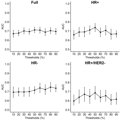 |
Effect of enhancement segmentation thresholds on predicting neoadjuvant response in breast cancer patients using DCE-MRI textural features
Deep K Hathi1, Rohan Nadkarni1, Natsuko Onishi1, Alex Anh-Tu Nguyen1, Wen Li1, Efstathios D Gennatas2, Bonnie N Joe1, Elissa R Price1, I-SPY 2 Consortium3, David C Newitt1, Ella F Jones1, and Nola M Hylton1
1Radiology and Biomedical Imaging, University of California San Francisco, San Francisco, CA, United States, 2Epidemiology & Biostatistics, University of California San Francisco, San Francisco, CA, United States, 3Quantum Leap Healthcare Collaborative, San Francisco, CA, United States
This study explores the prediction of pathologic complete response (pCR) using tumor-derived textural features in breast cancer patients receiving neoadjuvant chemotherapy. Textural features were generated from increasingly restricted tumor masks applied on DCE-MRI signal enhancement ratio maps. Elastic net and random forests models were trained on features from baseline and early treatment timepoints, resulting in minimal differences in AUC between percent enhancement segmentation thresholds and a mean AUC of 0.68 (range 0.60-0.75). Our analysis suggests that, for the prediction of pCR, textural features derived from strongly enhancing regions dominate over those from regions of lower enhancement.
|
|
0144.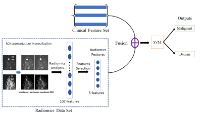 |
Predicting malignancy in additional lesion in breast cancer: A machine learning approach combining radiomics and clinical imaging analysis
Tien Anh Nguyen1, Hyo Jae Lee2, Luu-Ngoc Do1, Hyo-Soon Lim2,3, and Ilwoo Park3,4,5
1Radiology, Chonnam National University, GWANGJU, Korea, Republic of, 2Radiology, Chonnam National University Hwasun Hospital, Hwasun, Korea, Republic of, 3Radiology, Chonnam National University, Gwangju, Korea, Republic of, 4Radiology, Chonnam National University Hospital, GWANGJU, Korea, Republic of, 5Artificial Intelligence Convergence, Chonnam National University, Gwangju, Korea, Republic of
The purpose of this study was to investigate the feasibility of machine learning classifiers combining radiomics and clinical imaging interpretation for predicting malignancy in additional MR-detected enhancing lesions on multiparametric breast MRI. Machine learning algorithms trained with the combination of radiomics features extracted from breast MRI and clinical imaging interpretation what was obtained by an experienced breast radiologist demonstrated the maximal accuracy and AUC of 86.2% and 92.6%, respectively. The results from this study suggest that our approach may provide a noninvasive assisting tool to guide proper management that can reduce the use of unnecessary US or biopsy.
|
The International Society for Magnetic Resonance in Medicine is accredited by the Accreditation Council for Continuing Medical Education to provide continuing medical education for physicians.
 Watch the Video