Combined Educational & Scientific Session
SARS-CoV-2: What We Know, Lessons Learned & Where We May Be Headed: When SARS-CoV-2 Attacks
ISMRM & SMRT Annual Meeting • 15-20 May 2021

Combined Educational & Scientific Session
SARS-CoV-2: What We Know, Lessons Learned & Where We May Be Headed: When SARS-CoV-2 Attacks
| Concurrent 6 | 18:00 - 20:00 | Moderators: Meiyun Wang & Christopher Filippi |
 |
When SARS-CoV-2 Attacks (Pathophysiology/Immunology/Vaccine)
Amit Mahajan
COVID-19 has created havoc in communities around the world, ever since the start of the pandemic in December 2019. The causative agent, SARS-CoV-2 is a novel Coronavirus, that produces a diversity of manifestations ranging from asymptomatic infections to fatal infections characterized by viral pneumonia, ARDS, systemic inflammation and thromboembolism. The current presentation aims to summarize the pathogenesis and immunology of COVID-19 and its impact on vaccinations and the newly discovered viral variants.
|
|
 |
Brain MR & SARS-CoV-2: Imaging Findings, Complications & Outcomes
Simonetta Gerevini
A relevant number of COVID-19 patients may present with neurological symptoms in the acute/subacute phase of the disease. Neuroimaging can reveal a wide spectrum of CNS abnormalities, from ischemic lesions to microhemorrhages as well as meningoencephalitis and extensive white matters lesions. Advanced imaging techniques (DWI, DTI, f-MRI) may reveal underlining “inflammation of the brain” in case of persistent neurological symptoms and an unremarkable MRI examination.Long term neurological and neuropsychological sequelae are reported up to 30–40% in COVID-19 survivors, includingfatigue, myalgia, headache, dysautonomia and cognitive impairment (“brain fog”). A complete understanding of these manifestations is mandatory.
|
|
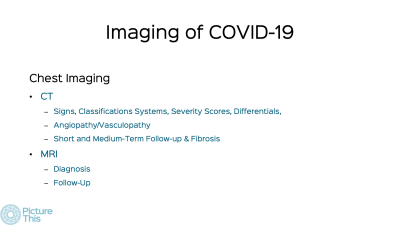 |
SARS-CoV-2 in the Lungs & the Role of MRI
Bhavin Jankharia
Imaging has an important role to play in the diagnosis, management and follow-up of patients with Covid-19. CT scan is an important modality. There are typical findings that allow a diagnosis of Covid-19 with high specificity. There are some unique signs due to the presence of angiopathy such as vascular "tree-in-bud" and target sign and perfusion defects without thrombosis. Follow-up of patients in the medium term usually shows regression of lesions. Some show persistent perfusion defects. MRI may show Covid-19 findings but cannot used routinely. Hyperpolarized Xe and O2 enhanced UTE MRI are being explored as options for follow-up. |
|
 |
Emerging MRI Data & Controversies of Cardiac Complications Related to SARS-CoV-2
Yuchi Han
There is increasing data describing abnormalities present on patients who have had CMR post SARS-CoV2 infection, especially in patients with troponin elevation and/or cardiac symptoms. These findings include infarct and myocarditis pattern late gadolinium enhancement, elevated T1, T2, and extracellular fraction levels. Controversy exists regarding whether or not these findings can be attributed to SARS-CoV2. Thus, we began by describing the definitions of myocarditis, the updated Lake Louise criteria for CMR for myocardial inflammation, and autopsy evidence of myocarditis associated with SARS-CoV2, and then go into the findings in the current literature.
|
| Concurrent 6 | 18:00 - 20:00 | Moderators: |
0215.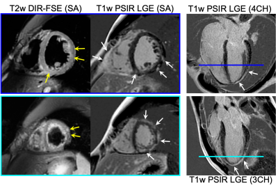 |
Evaluation for Myocarditis in Competitive Athletes Recovering from COVID-19 using Cardiac MRI
Jitka Starekova1, David A Bluemke1,2, William S Bradham1,2, Lee L Eckhardt2, Thomas M Grist1,3,4, Joanna E Kusmirek1, Christopher S Purtell2, Mark L Schiebler1, and Scott B Reeder1,2,3,4,5
1Radiology, University of Wisconsin-Madison, Madison, WI, United States, 2Medicine, University of Wisconsin-Madison, Madison, WI, United States, 3Biomedical Engineering, University of Wisconsin-Madison, Madison, WI, United States, 4Medical Physics, University of Wisconsin-Madison, Madison, WI, United States, 5Emergency Medicine, University of Wisconsin-Madison, Madison, WI, United States
Recent reports of COVID-19-associated myocarditis have raised safety concerns for athletes returning to training after recovering from COVID-19. As a result, our institution initiated a comprehensive screening program for all student athletes recovering from COVID-19, to screen for myocarditis using non-invasive diagnostic tests, including cardiac MRI. In this retrospective study we describe our institutional experience, including the prevalence and severity of MRI findings of myocarditis in student athletes recovering from COVID-19. Patients had mild (73/149;49%) or moderate symptoms (40/149;27%), or were asymptomatic (26/149;17%). Only 2/149 patients had MRI findings consistent with updated Lake Louise criteria for myocarditis (1.3%, 95%CI 0.4%,4.8%).
|
||
0216.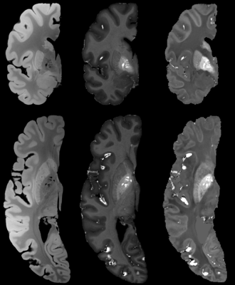 |
Ex vivo quantitative MR characterization of brain lesions induced by SARS-COV-2 infection
Mathieu David Santin1,2, Isabelle Plu3, Lydia Chougar1,2,3, Nadya Pyatigorskaya1,2,3, Roberto Toro4, Stéphane Lehéricy1,2,3, and Danielle Seilhean2,3
1Center for NeuroImaging Research - CENIR, Paris Brain Institute - ICM, Paris, France, 2ICM, Sorbonne University, UPMC Univ Paris 06, Inserm U1127, CNRS UMR 7225, Paris, France, 3Hôpital Pitié-Salpêtrière, AP-HP, Paris, France, 4Institut Pasteur, Paris, France
Our study aims to link ex vivo brain MRI signal abnormalities with neuropathological findings relative to the SARS-CoV-2 infection. MRI offers a “big picture” image on the whole organ compared to histology alone, which can be limited to blindly sampled small sections, when recent imagery is not available. The objective is to characterize the brain lesions linked to the viral infection. Our project should produce a new description of the anatomical structures affected by the infection in the central nervous system, and in particular those related to the brain vascular system.
|
||
 |
0217.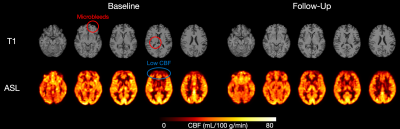 |
Examining cerebral blood flow in adults with COVID-19
William S.H. Kim1,2, Xiang Ji2, J. Jean Chen1,3, Asaf Gilboa3, Eugenie Roudaia3, Allison Sekuler3, Aravinthan Jegatheesan4, Mario Masellis2, Benjamin Lam2, Robert Fowler5, Chris Heyn2, Sandra E. Black2, Simon J. Graham1,4,
and Bradley J. MacIntosh1,2
1Department of Medical Biophysics, University of Toronto, Toronto, ON, Canada, 2Hurvitz Brain Sciences Program, Sunnybrook Research Institute, Toronto, ON, Canada, 3Rotman Research Institute, Toronto, ON, Canada, 4Physical Sciences Platform, Sunnybrook Research Institute, Toronto, ON, Canada, 5Evaluative Clinical Sciences, Trauma, Emergency & Critical Care, Sunnybrook Research Institute, Toronto, ON, Canada
The relationship between coronavirus disease 2019 (COVID-19) and cerebral blood flow (CBF) is not well understood. Here, we report on CBF measured by pseudo-continuous arterial spin labeling among adults that weeks prior had experienced flu-like symptoms with either a positive or negative COVID-19 diagnosis. Recruitment is ongoing, but at present we report no group differences in CBF across brain grey matter. However, subsequent regional analyses point to possible CBF abnormalities in those with a positive COVID-19 diagnosis.
|
|
|
0218.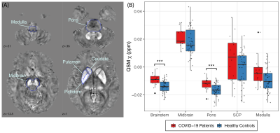 |
Characterization of brain susceptibility changes in post-hospitalisation COVID-19 patients at 7 Tesla
Catarina Rua1,2, Christopher T Rodgers2,3, Virginia F J Newcombe2,4, Anne Manktelow4, Doris A Chatfield4, Stephen J Sawcer3, Joanne G Outtrim4, Victoria C Lupson2, Emmanuel A Stamatakis2,3,4, Guy B Williams2,3, William T Clarke5, Karen D Ersche6,7, Kyle Pattinson5,
Edward T Bullmore6,7, David K Menon2,4,8, and James B Rowe1,9
1Department of Clinical Neurosciences and University of Cambridge Centre for Parkinson-plus, University of Cambridge, Cambridge, United Kingdom, 2Wolfson Brain Imaging Centre, University of Cambridge, Cambridge, United Kingdom, 3Department of Clinical Neurosciences, University of Cambridge, Cambridge, United Kingdom, 4Division of Anaesthesia, University of Cambridge, Cambridge, United Kingdom, 5Welcome Centre for Integrative Neuroimaging (FMRIB), University of Oxford, Oxford, United Kingdom, 6Department of Psychiatry, University of Cambridge, Cambridge, United Kingdom, 7Behavioural and Clinical Neuroscience Institute, University of Cambridge, Cambridge, United Kingdom, 8on behalf of the Cambridge NeuroCOVID group (www.wbic.cam.ac.uk/neuro-covid/) and the CITIID-NIHR COVID-19 BioResource Collaboration, Cambridge, United Kingdom, 9Medical Research Council Cognition and Brain Sciences Unit, University of Cambridge, Cambridge, United Kingdom
Patients hospitalized with the new coronavirus disease (COVID-19) have shown severe changes in the central nervous system (CNS), particularly microhaemorrhages and encephalitis. However, long-term effects on the CNS haven not yet been fully characterised. In this study we scanned a group of 14 recently hospitalized COVID-19 patients at ultra-high field (7T) and analysed the microstructural changes measured by quantitative susceptibility mapping (QSM) from both subcortical nuclei and brainstem, which are thought to be targeted by the virus.
|
|
0219.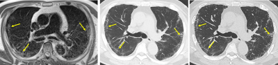 |
Low-Field Non-Contrast Cardiopulmonary MRI for Morphologic and Functional Assessment in Post-Covid Patients
Lea Azour1, William H Moore1, Larry Latson1, Mary Bruno1, Mahesh Bharath Keerthivasan2, Rany Condos3, Derek Mason1, Anna Shmukler1, Terlika Sood1, Adrienne Campbell-Washburn4, and Hersh Chandarana1
1Radiology, NYU Langone Health, New York, NY, United States, 2Siemens Medical Solutions, New York, NY, United States, 3Pulmonary Medicine, NYU Langone Health, New York, NY, United States, 4National Heart, Lung and Blood Institute, National Institutes of Health, Bethesda, MD, United States
Low-field (.55T) non-contrast MRI was performed in 15 post-Covid patients for combined cardiac and pulmonary evaluation, allowing for derivation of cardiac function in terms of left ventricular ejection fraction, as well as assessment for presence of persisting pulmonary parenchymal abnormalities. As the number of post-Covid patients increases, a radiation and contrast-free mode of cardiopulmonary imaging is of increasing relevance; low-field MRI in particular is a promising tool for high-performance lung imaging.
|
||
0220.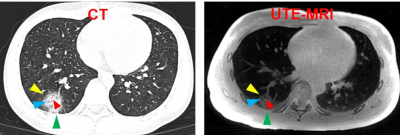 |
Assessing pulmonary findings of COVID-19 with ultrashort TE magnetic resonance imaging
Shuyi Yang1, Yunfei Zhang2, Fei Shan1, and Yongming Dai2
1Shanghai Public Health Clinical Center, Shanghai, China, 2Central Research Institute, United Imaging Healthcare, Shanghai, China
Chest computed tomography (CT) has played significant role in assessing the pulmonary findings of COVID-19. However, MRI, a radiation-free imaging technique has hardly been applied for assessing the pulmonary abnormalities of patients with COVID-19. Ultra shot echo time MRI (UTE-MR) is broadly regarded as a valuable tool for pulmonary imaging. This research, hence, aims to evaluate the COVID-19 with UTE-MR. The results indicated that there was no significant difference of image quality between the UTE-MR and CT for identifying the representative abnormalities. Besides, the intermethod agreement of UTE-MR and CT for assessing the pulmonary abnormalities were all determined excellent.
|
The International Society for Magnetic Resonance in Medicine is accredited by the Accreditation Council for Continuing Medical Education to provide continuing medical education for physicians.
 Watch the Video