Combined Educational & Scientific Session
Improved Body Imaging: Biliary, Liver & Pancreas
ISMRM & SMRT Annual Meeting • 15-20 May 2021

Combined Educational & Scientific Session
Improved Body Imaging: Biliary, Liver & Pancreas
| Concurrent 5 | 12:00 - 14:00 | Moderators: William Masch & Bin Song |
| Difficult Choices in Biliary Imaging: MRCP & Contrast Agents Video Permission Withheld
JeongHee Yoon
MRI including MRCP is a non-invasive modality to evaluate the bile duct.
|
||
 |
Differential Diagnosis in Biliary MRI Video Permission Withheld
Federica Vernuccio
Biliary MRI has a 95% sensitivity and 97% specificity for the detection of biliary dilatation. The purposes of this presentation are:– to describe imaging features of various obstructive and non-obstructive biliary diseases, providing tips for distinction between dilated bile duct pathologies;– to demonstrate the spectrum of imaging findings of focal or diffuse thickening of gallbladder wall and biliary cystadenoma/carcinoma;– to show other uncommon clinical scenarios where MRI may be useful to demonstrate biliary tree diseases.
|
|
| Cholangiocarcinoma: A Surgeon's Perspective
Christopher Sonnenday
|
||
| Case Discussion
Maxime Ronot
Selected challenging cases of biliary disease will be presented and discuss. Cases will illustrate the complex diagnostic process and work-up of patients with bile duct anomalies.
|
| Concurrent 5 | 12:00 - 14:00 | Moderators: |
0477. |
Retrospective Assessment of the Impact of Primary Language Video Instructions on Image Quality of Abdominal MRI
Myles Todd Taffel1, Andrew Rosenkrantz1, Jonathan Foster1, Jay Karajgikar1, Paul Smereka1, Thomas Mulholland1, Hoi Cheng Zhang1, Felicia Calasso1, Rebecca Anthopolos2, Kun Qian2, and Hersh Chandarana1
1Radiology, NYU Langone Medical Center, New York, NY, United States, 2NYU Langone Medical Center, New York, NY, United States
Previous work has demonstrated inferior abdominal MRI image quality in non-English speaking patients who require a translator. While a translator may be helpful, they are often interpreting remotely via a telephone and may be unfamiliar with the intricate MRI breathing instructions. As the result of the patient watching an instructional video explaining the MRI procedure in their primary language, image quality improves to match that of studies performed in primary English speaking patients.
|
||
 |
0478.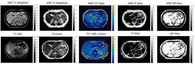 |
Free Breathing 2D Abdominal Magnetic Resonance Fingerprinting with quadratic RF phase
Sherry Huang1, Yong Chen2, Reid Bolding3, Leonardo Kayat Bittencourt4, Mark Griswold2, and Rasim Boyacioglu2
1Biomedical Engineering, Case Western Reserve University, Cleveland, OH, United States, 2Radiology, Case Western Reserve University, Cleveland, OH, United States, 3Physics, Case Western Reserve University, Cleveland, OH, United States, 4University Hospitals Cleveland Medical Center, Cleveland, OH, United States
This study presents a Pilot Tone (PT) based free-breathing technique for two-dimensional simultaneous quantification of T1, T2, T2*, fat fraction (FF), water fraction (WF), and off-resonance. This technique integrates quadratic RF phase-based Magnetic Resonance Fingerprinting (qRF-MRF) and PT navigator to retrospectively provide simultaneous quantification of multiple tissue properties in the abdomen at end-inhalation and end-expiration states.
|
|
0479.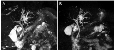 |
Breath-hold 3D gradient- and spin-echo (GRASE) MRCP compared to compressed-sensing highly accelerated respiratory-triggered technique.
Nobuyuki Kawai1, Yoshifumi Noda1, Kimihiro Kajita2, Hiroshi Kawada1, and Masayuki Matsuo1
1Radiology, Gifu University, Gifu, Japan, 2Radiology Services, Gifu University Hospital, Gifu, Japan
MR cholangiopancreatography (MRCP) plays an essential role in the noninvasive assessment of the biliary and pancreatic duct systems. Although the conventional respiratory-triggered three-dimensional turbo spin-echo (RT-3D-TSE) MRCP sequence has an excellent duct-to-periductal tissue contrast, the long acquisition time, over 5 minutes, has been a clinical burden. We assessed two types of ultrafast MRCP within 30 seconds of scan time. Breath-hold 3D gradient- and spin-echo (GRASE) MRCP provided better image quality and a reduced number of poor or non-diagnostic images compared to RT-3D-TSE MRCP highly accelerated with optimized integrated combination with parallel imaging and compressed-sensing technique (Compressed SENSE).
|
||
0480.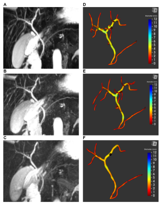 |
Comparison of Quantitative 3D Magnetic Resonance Cholangiopancreatography Biliary Tree Metrics derived from 3 Different Acquisition Methods
Neeraja Mahalingam1, George Ralli2, Gerard Ridgway2, Andrew Trout3,4,5, and Jonathan Dillman3,4,6
1Imaging Research Center, Cincinnati Children's Hospital Medical Center, Cincinnati, OH, United States, 2Perspectum Ltd., Oxford, United Kingdom, 3Department of Radiology, Cincinnati Children's Hospital Medical Center, Cincinnati, OH, United States, 4Department of Radiology, University of Cincinnati College of Medicine, Cincinnati, OH, United States, 5Department of Pediatrics, University of Cincinnati College of Medicine, Cincinnati, OH, United States, 6Center for Autoimmune Liver Disease, Cincinnati Children's Hospital Medical Center, Cincinnati, OH, United States
Comparison of the performance of different methods of acquiring three-dimensional (3D) magnetic resonance cholangiopancreatography (MRCP) data have been largely qualitative. MRCP+ prototype software (Perspectum Ltd.; Oxford, UK) was used to derive quantitative biliary tree metrics from 3D MRCP acquired using three different methods. Intra-class correlation coefficients (ICCs) demonstrated strong agreement on biliary tree volume, median intrahepatic duct diameters, number of ducts, and length of dilations between 3D FSE and CS-FSE MRCP (ICCs=0.84-0.93); there was slightly less agreement between CS-FSE and 3D GRASE MRCP. Our results suggest that CS-FSE provides comparable visualization of the biliary system to conventional 3D FSE MRCP.
|
||
0481.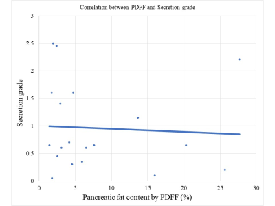 |
Investigation of risk factors for pancreatic exocrine insufficiency using 3T multiparametric MR imaging
Hidemitsu Sotozono1, Akihiko Kanki1, Kazuya Yasokawa1, Akira Yamamoto1, Tsutomu Tamada1, and Yu Ueda2
1Radiology, Kawasaki Medical School, Kurashiki, Japan, 2Philips Japan, Tokyo, Japan
Retrospective study included 46 patients without pancreatic disorders including pancreatic tumor and acute pancreatitis who underwent 3T abdominal multi-parametric MRI including cine-dynamic with spatially selective inversion recovery pulse and proton density fat fraction in 3D mDIXON quant (PDFF (%)). Pancreatic exocrine function was assessed as secretion grade (SG) based on the travel distance of inflowing pancreatic juice in the main pancreatic duct on cine-dynamic MRCP. The negative correlation between PDFF of the pancreas and SG estimated by cine-dynamic MRCP with spatially selective IR pulse suggests an association between pancreatic steatosis and impaired pancreatic exocrine function.
|
||
0482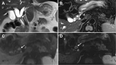 |
Hilar cholangiocarcinoma evaluation using zoomed echo-planar DW imaging with 2D spatial-selective radiofrequency excitation pulses Video Permission Withheld
Jingjing Liu1, Mengyue Huang1, Jingliang Cheng1, and Jinxia Zhu2
1Department of MR Imaging, the First Affiliated Hospital of Zhengzhou University, Zhengzhou, China, 2MR Collaboration, Siemens Healthcare Ltd, Beijing, China
We investigated the feasibility of using and the resultant image quality of zoomed diffusion-weighted echo-planar imaging (z-EPI) for hilar cholangiocarcinoma assessments. Compared with conventional single-shot EPI (c-EPI), z-EPI showed better delineation of anatomic structures in the hepatic hilar region, better lesion conspicuity, and overall higher image quality. Bile duct wall lesion delineations and lumens were also improved in z-EPI in four of 16 patients. These findings suggest that z-EPI may be preferred for improved imaging of hilar cholangiocarcinoma.
|
The International Society for Magnetic Resonance in Medicine is accredited by the Accreditation Council for Continuing Medical Education to provide continuing medical education for physicians.
 Watch the Video