Combined Educational & Scientific Session
Real-Time Cardiac MRI
ISMRM & SMRT Annual Meeting • 15-20 May 2021

Combined Educational & Scientific Session
Real-Time Cardiac MRI
| Concurrent 2 | 14:00 - 16:00 | Moderators: Holden Wu & Eugene Kholmovski |
 |
Technical Overview of Real-Time Cardiac MRI
Ganesh Adluru
Real-time cardiac MRI allows for the application of cardiac MRI to broader patient populations. Real-time CMR involves rapid data acquisition while free-breathing, and is typically independent of ECG-gating. Undersampled k-space acquisitions combined with advanced reconstruction methods are essential for real-time CMR. Compressed sensing (CS) is a popular approach that involves data sampling patterns that generate incoherent artifacts and uses iterative L1 norm constrained reconstructions. However, CS reconstructions are time-consuming. Deep-learning reconstructions offer real-time reconstructions from undersampled data as well as rapid automatic post-processing. The talk will give an overview of technical aspects for some of the real-time applications.
|
|
| Pediatric Applications of Real-Time Cardiac MRI
Vivek Muthurangu
In this talk the basics of real-time in paediatric clinical practice. I will discuss state-of-the-art compressed sensing reconstructions for assessment of ventricular function and blood flow using non-Cartesian acquisitions. I will also discuss the use of machine learning as an alternative to compressed sensing for faster reconstruction and easier clinical deployment. Finally, I will discuss the use of real-time for exercise MRI. |
||
| Real-Time Imaging During Exercise Stress Video Permission Withheld
Reza Nezafat
|
||
 |
Real-Time Cardiac MRI for MR-guided Interventions
Michael Bock
Real-time MRI to guide interventional cardiac procedures is challenging as images need to be acquired with high frame rates of 5-10 Hz, image reconstruction must be performed with low latency, and images need to have high signal-to-noise ratio. During the intervention different contrasts are needed to visualize the instruments and to assess the anatomical and functional changes. To navigate instruments such as catheters in the coronary vasculature, active tracking methods are beneficial, and interactive display software is needed to visualize and track the instruments at the patient table.
|
|
 |
Future Directions of Real-Time MRI
Adrienne Campbell-Washburn
This talk will describe recent developments in real-time cardiac MR (CMR) and predict some trends that will incorporated into real-time CMR in the future. This talk describes new acquisition methods relevant to real-time CMR, the role of machine learning, computational methods for low-latency image reconstruction, and the potential of real-time CMR at lower field strengths. Real-time CMR for diagnostic imaging, monitoring of physiological provocations and MRI-guided interventions are considered.
|
| Concurrent 2 | 14:00 - 16:00 | Moderators: |
 |
0503.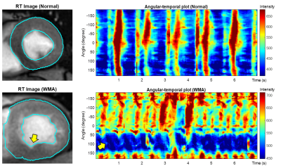 |
Rapid Physiological Dynamics Measured by Real-Time MRI at Up to 100Hz: MR Kinematography
Dan Zhu1, Tricia Steinberg2, Robert G. Weiss2, Dirk Voit3, Jens Frahm3, and Paul A. Bottomley4
1Department of Biomedical Engineering, Johns Hopkins University, Baltimore, MD, United States, 2The Division of Cardiology, Department of Medicine, Johns Hopkins University, Baltimore, MD, United States, 3Biomedizinische NMR, Max-Planck-Institut fur biophysikalische Chemie, Gottingen, Germany, 4The Division of MR Research, Department of Radiology, Johns Hopkins University, Baltimore, MD, United States
The advent of high-speed real-time (RT) MRI permits monitoring of physiological function at unprecedented frame-rates. Here, physiological dynamics at 25-100 frames-per-second are explored using temporal domain Fourier transform (FT) and principal component analysis (PCA). RT cerebral, cardiac and pharyngeal datasets are acquired with continuous radial encoding and nonlinear inverse reconstruction implemented in graphics processing units. FT detects spectral patterns in pharyngeal images acquired during speaking. FT and PCA reflect components associated with breathing and cardiac functions in the brain while decomposition and synthesis in the time-domain can pinpoint cardiac wall motion abnormalities in patients with heart disease.
|
|
0504.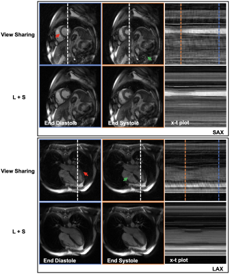 |
Spiral-in-out bSSFP Real-Time Cine on a High Performance 0.55T Scanner
Zhixing Wang1, Xue Feng1, John P. Mugler2, Michael Salerno3, Adrienne E. Campbell-Washburn4, and Craig H. Meyer1
1Biomedical Engineering, University of Virginia, Charlottesville, VA, United States, 2Radiology & Medical Imaging, University of Virginia, Charlottesville, VA, United States, 3School of Medicine, University of Virginia, Charlottesville, VA, United States, 4Cardiovascular Branch, Division of Intramural Research, National Heart, Lung, and Blood Institute, National Institutes of Health, Bethesda, MD, United States
This abstract describes a spiral-in-out bSSFP pulse sequence combined with a low rank plus sparse (L+S) image reconstruction for ungated real-time cine on a high performance 0.55T MRI scanner. Preliminary results show that the proposed method is a promising technique for real-time cardiac imaging with high image quality and excellent temporal resolution.
|
||
 |
0505.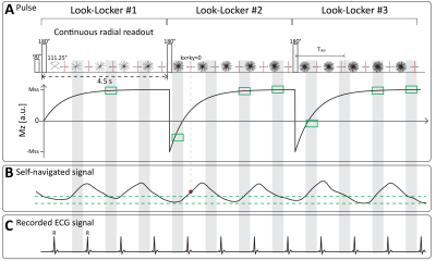 |
A free-running cardiac T1* mapping sequence for quantifying dynamic changes in myocardial T1* at rest and physiological exercise
Rui Guo1, Haikun Qi2, Xiaoying Cai1,3, Selcuk Kucukseymen1, Hassan Haji-Valizadeh1, Jennifer Rodriguez1, Amanda Paskavitz1, Patrick Pierce1, Beth Goddu1, Richard B. Thompson4, and Reza Nezafat1
1Department of Medicine (Cardiovascular Division), Beth Israel Deaconess Medical Center and Harvard Medical School, BOSTON, MA, United States, 2School of Biomedical Engineering and Imaging Sciences, London, United Kingdom, 3Siemens Medical Solutions USA, Inc, Boston, MA, United States, 4University of Alberta, Department of Biomedical Engineering, Edmonton, MA, Canada
In this study, we developed a free-running cardiac T1* mapping sequence (DELTA) for quantifying the dynamic changes in myocardial T1* in response to physiological exercise. DELTA adopts continuous radial acquisition, self-navigation, and adaptive acquisition window to address the challenges associated with high heart rate and deep breathing after exercise. Phantom T1* by DELTA was heart-rate insensitive and had good repeatability. In vivo T1* among multiple measurements had little variation and comparable precision with MOLLI5(3)3 T1*.In the stress/rest studies,
T1* reactivity was larger during the first scan after exercise and gradually reduced along the duration of post-exercise.
|
|
|
0506.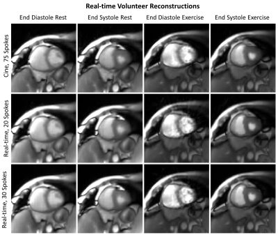 |
Real-time Cardiac MRI during Exercise with Radial Sampling and Compressed Sensing: Evaluation in a Numerical Phantom and In-Vivo
Philip A Corrado1, Daniel Seiter1, Christopher J François2, Farhan Raza1, Kevin Johnson1, and Oliver Wieben1
1University of Wisconsin-Madison, Madison, WI, United States, 2Mayo Clinic, Rochester, MN, United States
We used radial sampling, parallel imaging, and compressed sensing for real-time cardiac MRI during exercise, comparing the performance of two temporal resolutions for this approach in a numerical phantom and in a human volunteer. We found the approach feasible with sufficient spatial and temporal resolution to capture myocardial motion. While a longer temporal resolution with 30 radial spokes provides better image quality during rest, shortening the temporal resolution by acquiring just 20 spokes can improve results during exercise by better capturing rapid motion such as late diastolic filling.
|
|
0507.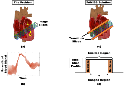 |
Flow and Motion Insensitive Steady State (FAMISS): Advancing ungated steady-state cardiac perfusion
Jason Mendes1, Johnathan Le1, Mark Ibrahim2, Ganesh Adluru1, and Edward DiBella1
1Radiology and Imaging Sciences, University Of Utah, Salt Lake City, UT, United States, 2Cardiovascular Medicine, University Of Utah, Salt Lake City, UT, United States
Quantitative cardiac perfusion requires reliable dynamic measurement of the contrast agent concentration in both myocardium and blood. Ungated methods offer immunity to poor ECG signals, acquire multiple cardiac phases and have been shown to achieve comparable contrast-to-noise as saturation recovery based methods. However, blood flow and cardiac motion can interrupt the steady-state and result in the calculation of inaccurate contrast agent concentrations. These errors in perfusion quantification can be avoided using a proposed Flow and Motion Insensitive Steady State (FAMISS) technique.
|
||
0508.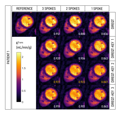 |
The key to extremely accelerated model-based quantitative first-pass perfusion cardiac MRI
Teresa M Correia1, Torben Schneider2, and Amedeo Chiribiri1
1School of Biomedical Engineering and Imaging Sciences, King's College London, London, United Kingdom, 2Philips Healthcare, Guildford, United Kingdom
First-pass perfusion cardiac MR (FP-CMR) is one of the methods of choice for evaluating myocardial ischemia. Moreover, quantitative FP-CMR methods that provide pixel-wise quantitative myocardial perfusion maps are increasingly being applied as an alternative to visual inspection. Recently, a DIRect QuanTitative (DIREQT) FP-CMR model-based reconstruction has been proposed to directly estimate myocardial perfusion maps and highly accelerate FP-CMR scans. Here, DIREQT is combined with the idea of view-sharing and KEYhole imaging (DIREQT-KEY) to improve DIREQT reconstructions, particularly from extremely accelerated FP-CMR acquisitions. DIREQT-KEY directly generates high-quality quantitative myocardial perfusion maps from less than 4 radial spokes per time frame.
|
The International Society for Magnetic Resonance in Medicine is accredited by the Accreditation Council for Continuing Medical Education to provide continuing medical education for physicians.
 Watch the Video