Combined Educational & Scientific Session
Emerging Quantitative Contrasts: QSM, ETPM & MRE
ISMRM & SMRT Annual Meeting • 15-20 May 2021

Combined Educational & Scientific Session
Emerging Quantitative Contrasts: QSM, ETPM & MRE
Jump to:
Quantitative Susceptibility Mapping (QSM)
Electrical Tissue Property Mapping
Magnetic Resonance Elastography (MRE)
QSM, ETPM & MRE
| Concurrent 2 | 14:00 - 16:00 | Moderators: Emma Biondetti |
Target Audience
This educational session is designed for researchers in both basic science and clinical research.
Educational Objectives
As a result of attending this course, participants should be able to:
- Explain the underpinning physics, contrast mechanisms, and reconstruction challenges of QSM;
- Describe the application of QSM in the brain as a biomarker of changes in tissue composition (e.g. myelin and iron) in neurodegenerative and neurovascular diseases; and
- Discuss future research directions in this area.
 |
QSM: Theory & Methods
Steffen Bollmann
This educational lecture will give a broad overview of Quantitative Susceptibility Mapping. After explaining what magnetic susceptibility is and how we can measure it, I cover the data acquisition aspects, coil combination, unwrapping, masking, background field correction and dipole inversion. Then I show different toolboxes that enable QSM processing. I discuss the topic of referencing QSM results in for example group studies and conclude with a bigger picture and wish list of features that I would like to see in the QSM method development going forward.
|
|
| QSM: Applications & Outlook
Sarah Eskreis-Winkler
|
||
 |
QSM: Applications & Outlook
Anita Karsa
Quantitative Susceptibility Mapping (QSM) calculates the tissue magnetic susceptibility map from the phase of gradient-echo MRI acquisitions. It provides new and complementary information to the magnitude at no extra cost of scanning time. However, implementing and optimising a suitable acquisition protocol and QSM processing pipeline is not trivial in practice. Here I discuss the practical aspects of performing QSM in clinical studies in and outside of the brain. I highlight common pitfalls and provide technical recommendations for beginners. Finally, I also discuss my outlook on the potential role of QSM in clinical practice.
|
| Concurrent 2 | 14:00 - 16:00 | Moderators: Xiaotong Zhang & Wyger Brink |
 |
Electrical Tissue Property Mapping: Theory & Methods
Rita Schmidt
We will discuss methods to map electrical properties (EP) – conductivity and permittivity - in MRI. The methods include MR Electrical Impedance Tomography (MREIT) and Electrical Properties Tomography (MREPT), each mapping EP in a different frequency range. The first study of MREPT by Haacke in 1991, relied on the Helmholtz-wave-based formulation, but allowed limited resolution. This led to several new methods, including gradient-based EPT (gEPT) and Contrast Source Inversion EPT (CSI-EPT). New studies incorporate deep learning and demonstrate high-resolution imaging of the brain. EPT has the potential of providing new biomarkers for a range of applications and for patient-specific energy estimation.
|
|
 |
Electrical Tissue Property Mapping: Applications & Outlook
Stefano Mandija
Tissue electrical properties represent an emerging quantitative MRI contrast. In this talk, we will review applications and outlook of MR-based Electrical Properties Tomography. We will briefly review applications of low frequencies (kHz) electrical properties mapping, with emphasis on guidance of brain stimulation. After, we will review applications of high frequencies (hundreds MHz) electrical properties mapping, with emphasis on tumor diagnosis. Finally, we will look at future directions, e.g. integration of deep learning solutions to resolve physics based reconstruction issues and application of Electrical Properties Tomography for early stage radiotherapy treatments response assessment.
|
| Concurrent 2 | 14:00 - 16:00 | Moderators: Yuan Feng & Dieter Klatt |
 |
MR Elastography: Theory & Methods
Meng Yin
Many disease processes cause marked changes in tissue mechanical properties. Mechanical properties are therefore promising biomarkers for monitoring and characterizing various pathophysiologic conditions of tissues. MR-based elastography is a phase-contrast technique for estimating the shear stiffness of tissues by imaging propagating shear waves generated from a standardized extrinsic vibrating source. In patient care, the innovative stiffness biomarker is beginning to see widespread clinical use for assessing hepatic fibrosis as an alternative to biopsy. There is significant potential to further develop elastography techniques to implement three-dimensional multiparametric methods that have promise for distinguishing varying pathophysiologic states of the entire organ.
|
|
 |
MR Elastography: Applications & Outlook
Philip Bayly
Magnetic resonance elastography (MRE) is a method to estimate the mechanical properties of soft tissue from MR images of vibration-induced shear waves. In this presentation, I introduce the fundamental principles of MRE: material properties and their relationships to wave speed; how to induce waves in tissue; basic imaging sequences used in MRE; and the inversion of shear wave fields to estimate mechanical properties. Applications of MRE include the clinically important, non-invasive measurement of liver stiffness, and emerging studies of brain, breast, kidney, intervertebral disc, and muscle. New MRE methods are under development to estimate anisotropic (direction-dependent) mechanical properties in tissue.
|
| Concurrent 2 | 14:00 - 16:00 | Moderators: |
 |
0735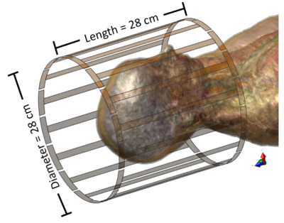 |
Neural network-based cr-EPT stabilization. Video Permission Withheld
Adan Jafet Garcia Inda1, Shao Ying Huang2,3, Nevrez Imamoglu4, and Wenwei Yu5
1Medical Engineering, Chiba University, Chiba, Japan, 2Department of Surgery, National University of Singapore, Singapore, Singapore, 3Engineering Product Development, Singapore University of Technology and Design, Singapore, Singapore, 4Artificial Intelligence Research Center, National Institute of Advanced Industrial Science and Technology, Tokyo, Japan, 5Center for Frontier Medical Engineering, Chiba University, Chiba, Japan
Electrical properties are a novel contrast mechanism for quantitative MRI. Conductivity can be used as a biomarker for tumorous tissues. Different analytic Magnetic-Resonance Electrical Properties Tomography (MREPT) methods have been proposed, however, accurate reconstructions require empirical assessment and setting of regularization coefficients per sample. In this work, based on a modified formulation of Convection-Reaction Equation-Based EPT (cr-EPT), the regularization coefficients are learned from the difference between reconstructed conductivity maps and their ground truth, using a convolutional neural network (CNN) model. The CNN model with the modified cr-EPT could achieve conductivity reconstructions with higher accuracy, compared to several analytical models.
|
|
0736.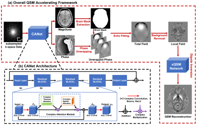 |
Accelerating QSM using Compressed Sensing and Deep Neural Network
Yang Gao1, Feng Liu1, Stuart Crozier1, and Hongfu Sun1
1The University of Queensland, Brisbane, Australia
Quantitative susceptibility mapping (QSM) has shown significant clinical potential for studying neurological disorders, but its acquisitions are relatively slow, e.g. 5-10 mins. Compressed sensing (CS) undersampling and reconstruction techniques have been used to accelerate the magnitude-based MRI acquisitions; however, most of them are ineffective to phase signal due to its non-convex nature. In this study, we propose a deep neural network “CANet” using complex attention modules to recover both the magnitude and phase images from the CS-undersampled data, enabling substantial acceleration of phase-based QSM.
|
||
0737.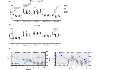 |
Brain stiffness changes before disease onset and reflects remission and relapse in a mouse model of multiple sclerosis
Anna Morr1, Rafaela Vieira da Silva1, Gergely Bertalan1, Stefan Paul Koch1, Susanne Mueller1, Philipp Boehm-Sturm1, Jürgen Braun1, Carmen Infante Duarte1, and Ingolf Sack1
1Charité - Universitätsmedizin Berlin, Berlin, Germany
The disease progression in a mouse model of multiple sclerosis (EAE) was monitored based on regionally resolved brain viscoelasticity. Multifrequency MR elastography was applied prior to immunization and during subsequent phases of disease progression. Our results suggest that mechanical structures of the brain, particularly throughout the whole brain, in the cortex, and in the periventricular and hippocampal areas, are already significantly affected by neuroinflammation bevor clinical disability is manifested. Moreover, stiffness of the hippocampus and periventricular tissue transiently recovered during remission and became re-affected during relapse. This suggests that mechanical alterations are transient and recover during remission.
|
||
0738.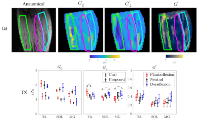 |
Validation and Application of a Novel Anisotropic MRE Reconstruction Method
Renee Miller1, Rob Lloyd2, Daniel Fovargue1, Behzad Babei2, Lauriane Juge2,3, Ralph Sinkus1, David Nordsletten1,4, and Lynne Bilston2,3
1Biomedical Engineering and Imaging Sciences, Kings College London, London, United Kingdom, 2Neuroscience Research Australia, Sydney, Australia, 3Faculty of Medicine, University of New South Wales, Sydney, Australia, 4University of Michigan, Ann Arbor, MI, United States
Due to their structure and composition, many biological tissues have been shown to be anisotropic. MRE, a non-invasive tool used to measure the stiffness of tissue, has previously been used to diagnose and track disease progression based on tissue stiffness changes. This work presents a robust and novel method for estimating anisotropic stiffness utilising harmonic displacements from MRE and material orientations from DTI. The method was verified using in silico experiments and applied to in vivo imaging data of the lower leg muscles in volunteers. In vivo stiffness estimates show changes in anisotropic stiffness with changes in passive muscle stretch.
|
||
|
0739.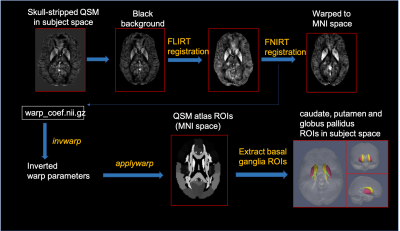 |
Relationships Between 7T Basal Ganglia QSM, Volume and Clinical Manifestation of Huntington’s Disease
Sivakami Avadiappan1, Melanie A Morrison1, Yicheng Chen1, Angela Jakary1, Nancy Cai2, Megan Casey2, Julia Glueck2, Joseph Tallakson2, Michael Geschwind2, Katherine Possin2, Alexandra Nelson2, Christopher P Hess1,2, and Janine M Lupo1
1Department of Radiology and Biomedical Imaging, University of California San Francisco, San Francisco, CA, United States, 2Department of Neurology, University of California San Francisco, San Francisco, CA, United States
Huntington’s Disease (HD) is a neurodegenerative disorder with severe cognitive and motor impairment caused by abnormal cytosine-adenine-guanine (CAG) repeat expansion within the HTT gene. The goal of this study was to explore differences in iron deposition measured by quantitative susceptibility mapping (QSM) and volume of basal ganglia structures between healthy controls and patients with pre-manifest HD or early manifest HD and correlate them with clinical variables. The caudate and putamen exhibited atrophy that increased with disease severity, CAP score, and impaired motor function. Iron deposition increased with the onset of symptoms, CAP scores, and cognitive decline.
|
|
0740.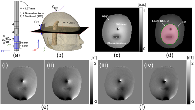 |
Imaging of electromagnetic field distribution in deep brain stimulation (DBS): a biological tissue phantom study
Munish Chauhan1, Saurav ZK Sajib1, Sulagna Sahu1, Enock Boakye1, Willard S Kasoff2, and Rosalind J Sadleir1
1School of Biological and Health System Engineering, Arizona State University, Tempe, AZ, United States, 2Department of Surgery, University of Arizona, Tucson, AZ, United States
Knowledge of electromagnetic field distribution parameters such as current density, conductivity, and electric field distribution may play an essential role in understanding DBS therapeutic effects on living tissues. However, until now there has been no experimental method that can measure these field parameters. In this work, we demonstrate that it is possible to quantify electromagnetic field distribution during DBS therapy using magnetic resonance electrical impedance tomography (MREIT).
|
The International Society for Magnetic Resonance in Medicine is accredited by the Accreditation Council for Continuing Medical Education to provide continuing medical education for physicians.
 Watch the Video