Combined Educational & Scientific Session
Interventional & Low-Field MRI
ISMRM & SMRT Annual Meeting • 15-20 May 2021

Combined Educational & Scientific Session
Interventional & Low-Field MRI
| Concurrent 3 | 16:00 - 18:00 | Moderators: Elena Kaye & Adrienne Campbell-Washburn |
 |
Rapid Imaging for MRI-Guided Interventions
Steven Allen
Rapid imaging techniques can improve patient outcomes and reduce costs during MRI-guided interventions. This talk will cover 5 common strategies for accelerating guidance MR acquisitions and give examples of their implementation from interventional MRI programs groups across the globe. These strategies include using time wisely, reducing fields of view, multiplexing, changing k-space trajectories, skipping data. I conclude with some brief speculation on the potential use of machine learning and AI to further accelerate guidance imaging.
|
|
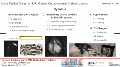 |
Active Device Design for MRI-Guided Cardiovascular Catheterizations
Ali Özen
In this lecture, active device design for cardiovascular interventions will be covered from a technical and a practical perspective. First, various RF coil designs and their working principles will be reviewed. Second, approaches to interface the active devices to the MRI system will be introduced. Afterwards, application of active devices from imaging to tracking will be presented. Finally, challenges and recent advancements in production, and MR safety of the active devices will be discussed.
|
|
| Hardware for MRI-Guided High Intensity Focused Ultrasound
Allison Payne
MRI guided focused ultrasound combines two innovative technologies to alter tissues non-invasively. The purpose of this talk is to describe the specialized hardware required by the MRI scanner to enable these MR guided focused ultrasound procedures to be performed in the clinical MR environment. While these hardware requirements are typically treatment site specific to some degree, there are key points that can be applied to any MR guided focused ultrasound system. This talk will address three key hardware aspects, specifically the effects of the ultrasound transducer on MR imaging, radiofrequency coils, and acoustically coupling the ultrasound transducer to the patient.
|
||
| Case Demonstration from an Interventional MRI Suite Video Permission Withheld
Sherif Nour
|
||
 |
MRI-Guided Robotic Prostate Biopsy
Junichi Tokuda
This presentation will overview the technologies and clinical application of MRI-guided robotic prostate biopsy. Robotic devices have been developed and clinically tested in recent years to address technical challenges in MRI-guided prostate biopsy, such as the limited access to the patient in the bore, and suboptimal needle placement accuracy. I will discuss the technologies for MR-guided robotic biopsy from several key aspects, including system architecture, MRI compatibility, kinematics design, software, imaging sequences, and evaluation of needle placement accuracy. Additionally, I will review some of the published studies that have demonstrated the clinical feasibility of MRI-guided robotic prostate biopsy.
|
| Concurrent 3 | 16:00 - 18:00 | Moderators: |
0797.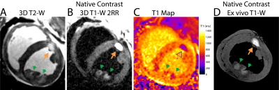 |
Native Contrast Visualization of Myocardial Radiofrequency Ablation and Acetic Acid Chemoablation Lesions at 0.55 T
Daniel Herzka1, Chris G. Bruce1, Rajiv Ramasawmy1, D. Korel A Yildirim1, Kendall J. O'Brien1, William H. Schenke1, Toby Rogers1,2, Adrienne E. Campbell-Washburn1,3, Robert J. Lederman 1, and Aravindan Kolandaivelu1,4
1NHLBI, Division of Intramural Research, National Institutes of Health, Bethesda, MD, United States, 2Department of Cardiology, Medstar Washington Hospital Center, Washington, DC, United States, 3Biophysics and Biochemistry Branch, Division of Intramural Research, National Institutes of Health, Bethesda, MD, United States, 4Division of Cardiology, Department of Medicine, Johns Hopkins University School of Medicine, Baltimore, MD, United States
This work determined feasibility of visualizing cardiac radiofrequency (RF) ablation lesions at low field (0.55 T) as well as a novel alternative method for targeted tissue destruction: acetic acid chemoablation. Native contrast T1-W imaging and T1 mapping and were carried out in vivo in swine on the day of ablation. Ex vivo high-resolution imaging and histology were used as references. T1 drop was higher for chemoablation (40%) than for RF ablation (19%) relative to myocardium, resulting in significantly higher SNR and CNRs. The visualization of coagulation necrosis from cardiac ablation is feasible using native-contrast low-field MRI.
|
||
0798.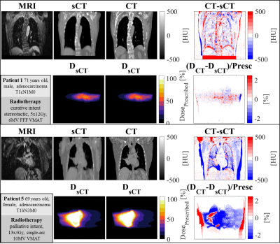 |
Deep learning-based 4D synthetic CT for lung radiotherapy
Matteo Maspero1,2, Kirsten M. Kerkering2,3, Tom Bruijnen1,2, Mark H. F. Savenije1,2, Joost J. C. Verhoeff1, Christoph Kolbitsch3, and Cornelis A. T. van den Berg1,2
1Radiotherapy, Division of Imaging & Oncology, UMC Utrecht, Utrecht, Netherlands, 2Center for Computational imaging group for MR diagnostic & therapy, Center for Image Sciences, UMC Utrecht, Utrecht, Netherlands, 3Physikalisch-Technische Bundesanstalt (PTB), Braunschweig and Berlin, Germany
The feasibility of generating synthetic CT for lung tumours from 4D MRI was investigated. A combination of multi-view 2D networks proved to be robust against image artefact and generated sCTs that enabled dose calculation on midposition sCTs. The proposed approach facilitates adaptive MR-guided radiotherapy reducing the time from patient positioning to irradiation and enables quality assurance with dose accumulation based on 4D MRI.
|
||
0799.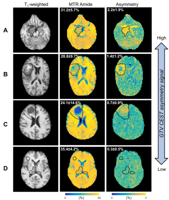 |
A Daily Quantitative Brain MRI Protocol for the 1.5 T MR-Linac: Feasibility of CEST with Preliminary Results on a Prospective Imaging Study
Rachel W. Chan1, Liam S.P. Lawrence2, Ryan T. Oglesby2, Hanbo Chen3, Brian Keller3, James Stewart3, Mark Ruschin3, Brige Chugh3,4, Scott MacKenzie3, Mikki Campbell3, Aimee Theriault3, Sten Myrehaug3, Jay Detsky3, Pejman J.
Maralani5, Chia-Lin Tseng3, Gregory J. Czarnota1,2,3, Greg J. Stanisz1,2,6, Arjun Sahgal3, and Angus Z. Lau1,2
1Physical Sciences, Sunnybrook Research Institute, Toronto, ON, Canada, 2Medical Biophysics, Sunnybrook Research Institute, Toronto, ON, Canada, 3Department of Radiation Oncology, Sunnybrook Health Sciences Centre, Toronto, ON, Canada, 4Department of Physics, Ryerson University, Toronto, ON, Canada, 5University of Toronto, Sunnybrook Health Sciences Centre, Medical Imaging, Toronto, ON, Canada, 6Department of Neurosurgery and Pediatric Neurosurgery, Medical University, Lublin, Poland
A multi-parametric imaging protocol, for monitoring patients with brain tumors treated using the 1.5T MR-Linac radiotherapy system, is presented with a focus on CEST. Phantom experiments were performed on the MR-Linac using varying concentrations of ammonium chloride mixtures. 24 subjects were included in the analysis. Mixed modelling was used to determine any differences between the gross tumor volume (GTV) and contralateral normal appearing white matter (cNAWM) regions, where three CEST parameters were investigated (MTRAmide, MTRNOE and Asymmetry). Results from phantom experiments confirmed detectable CEST asymmetry. Significant differences were found in all three CEST parameters between the GTV and cNAWM.
|
||
0800.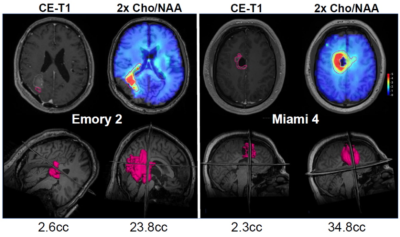 |
Spectroscopic MRI Guided Radiation Dose Escalation in Glioblastoma Patients
Mohammed Z Goryawala1, Eric A Mellon2, Saumya Gurbani3, Karthik Ramesh3, Brent D Weinberg4, Lawrence Kleinberg5, Eduard Schreibmann3, Sulaiman Sheriff1, Peter B. Barker6, Shu Hui-Kuo7, Hyunsuk Shim3, and Andrew Maudsley8
1Radiology, University of Miami School of Medicine, Miami, FL, United States, 2Radiation Oncology, University of Miami School of Medicine, Miami, FL, United States, 3Emory University, Atlanta, GA, United States, 4Radiology, Emory University, Atlanta, GA, United States, 5Radiation Oncology, Johns Hopkins University, Baltimore, MD, United States, 6Radiology, Johns Hopkins University, Baltimore, MD, United States, 7Radiation Oncology, Emory University, Atlanta, GA, United States, 8Radiology, University of Miami, School of Medicine, Miami, FL, United States
To improve localization of tumor tissue for radiation treatment planning a volumetric MR spectroscopic imaging acquisition was used to identify regions of metabolic abnormality. These regions were then integrated into the clinical treatment plan to target increased radiation dose for the identified volume. In a 3-site clinical trial of 30 patients with newly-diagnosed glioblastoma, the escalated dose resulted in a median overall survival of 23 months, which compares favorably with standard of care, without severe adverse events. This study demonstrates considerable potential for incorporating volumetric spectroscopic imaging into radiation treatment planning protocols.
|
||
 |
0801.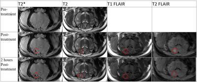 |
Technical feasibility and imaging of transcranial MR-guided in-vivo Histotripsy treatment
Dinank Gupta1, Ning Lu1, Adam Fox1, Dave Choi1, Jonathan Sukovich1, Badih Junior Daou2, Aditya Pandey2, Timothy Hall1, Zhen Xu1, and Douglas Noll1
1Biomedical Engineering, The University of Michigan, Ann Arbor, MI, United States, 2Department of Neurosurgery, The University of Michigan, Ann Arbor, MI, United States
Feasibility of transcranial MR-guided histotripsy for an in-vivo large animal model is established. Eight juvenile pigs were treated using an in-house, 128 channel histotripsy array at a wide range of sites around the brain. Target localization was done using fiducials placed on the array using pre-treatment MR images. Lesions were generated in all the pigs and were visualized in post treatment MRI scans and histology. In acute treatment phase, treatment effects were visible on T2*,T2, T1/T2 FLAIR weighted images. This is the first study to exhibit feasibility of in-vivo transcranial MR-guided histotripsy treatment.
|
|
0802.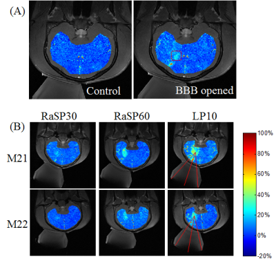 |
MR-Guided Blood-brain Barrier Opening Induced by Rapid Short-pulse Ultrasound on Non-human Primates
Hui Zhou1,2, Yang Liu3, Xiaojing Long3, Yangzi Qiao1, Jo Lee1, Chao Zou1,4, Xin Liu1,4, and Hairong Zheng1
1Lauterbur Imaging Research Center, Shenzhen Institutes of Advanced Technology, Chinese Academy of Sciences, Shenzhen, China, 2The Shenzhen College of Advanced Technology, University of Chinese Academy of Sciences, shenzhen, China, 3Research center for medical AI,Shenzhen Institutes of Advanced Technology, Chinese Academy of Sciences, Shenzhen, China, 4Key Laboratory for Magnetic Resonance and Multimodality Imaging of Guangdong Province, Shenzhen, China
The BBB has been opened with millisecond ultrasound in kinds of animals for researching of neurological diseases therapy. Rapid short-pulse (RaSP) ultrasound with a microsecond sequence has been proposed as a minimally disruptive and efficient method for BBB opening in mice and rats. This work quantitatively evaluate the feasibility and safety of BBB opening in non-human primate with RaSP by contrast enhanced MRI. The relative signal enhancement in RaSP with 6% energy deposition reached more than 60% of that with 10 ms long pulse (LP), which shows that RaSP is a practical method for BBB opening in a large-animal model.
|
The International Society for Magnetic Resonance in Medicine is accredited by the Accreditation Council for Continuing Medical Education to provide continuing medical education for physicians.
 Watch the Video