Combined Educational & Scientific Session
Diffusion Tractography
ISMRM & SMRT Annual Meeting • 15-20 May 2021

Combined Educational & Scientific Session
Diffusion Tractography
| Concurrent 4 | 18:00 - 20:00 | Moderators: Simona Schiavi & Rafael Henriques |
| Data Preprocessing & Diffusion Modeling
Carlo Pierpaoli
In this talk we will provide an “historical” overview of various modelling approaches that have been proposed for inferring anatomy and connectivity of white matter fibers from diffusion MRI data. We will describe the foundations of methods for deterministic tractography, and probabilistic tractography that will be addressed in detail by the subsequent lecturers. We will also provide background information to introduce the lecture on “validation” of Diffusion MRI tractography. Proper data preprocessing, however, can effectively minimize potential sources of artifacts. We will examine various steps that are desirable in a diffusion MRI preprocessing pipeline.
|
||
| Deterministic Tractography
Flavio Dell'Acqua
|
||
| Probabilistic Tractography
J. Donald Tournier
Probabilistic tractography approaches account for the inherent, multiple sources of uncertainty that impact on the fibre tracking process. They aim to provide a more representative depiction of the range of connections that are consistent with the data. These approaches typically depend on the availability of the distribution of fibre orientations, from which statistical samples can be obtained and used in the streamline propagation process. It is important to note that probabilistic approaches do not in general provide estimates of the probability of a connection; they aim to depict the full range of likely connections that are consistent with the data.
|
||
| Tractography Validation
Kurt Schilling
|
| Concurrent 4 | 18:00 - 20:00 | Moderators: |
 |
0863.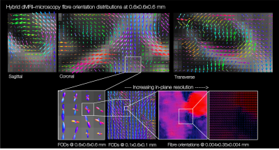 |
The microscopy connectome: towards 3D PLI tractography in the BigMac dataset
Amy FD Howard1, Istvan N Huszar1, Michiel Cottaar1, Greg Daubney2, Alexandre A Khrapitchev3, Rogier B Mars1,4, Jeroen Mollink1, Connor Scott5, Nicola Sibson3, Adele Smart1,5, Jerome Sallet2, Saad Jbabdi1, and Karla L Miller1
1FMRIB Centre, Wellcome Centre for Integrative Neuroimaging, University of Oxford, Oxford, United Kingdom, 2Wellcome Centre for Integrative Neuroimaging, Experimental Psychology, Medical Sciences Division, University of Oxford, Oxford, United Kingdom, 3MRC Oxford Institute for Radiation Oncology, Department of Oncology, University of Oxford, Oxford, United Kingdom, 4Donders Institute for Brain, Cognition and Behaviour, Radboud University Nijmegen, Nijmegen, Netherlands, 5Nuffield Department of Clinical Neurosciences, University of Oxford, Oxford, United Kingdom
The BigMac dataset is a unique resource that includes extensive MRI and densely sampled microscopy data acquired in a single, whole macaque brain. However, the high-resolution microscopy currently only informs on the fibre orientations in the 2D plane of sampled slides, precluding 3D reconstruction of the microscopy connectome. Here we use precise co-registration and joint modelling of diffusion MRI and polarised light images to reconstruct the microscopy fibre orientations in 3D. This will facilitate future determination of the whole brain, microscopy-inspired connectome, which we expect will provide neuroanatomical insight, and play a vital role in validating and advancing invivo tractography.
|
|
 |
0864.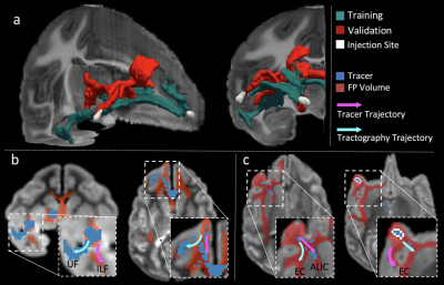 |
New insights from the IronTract challenge: Simple post-processing enhances the accuracy of diffusion tractography
Chiara Maffei1, Gabriel Girard2,3,4, Kurt G. Schilling5, Dogu Baran Aydogan6, Nagesh Adluru7, Andrey Zhylka8, Ye Wu9, Matteo Mancini10,11,12, Andac Hamamci13, Alessia Sarica14, Davood Karimi15, Fang-Cheng Yeh16, Mert E. Yildiz13,
Ali Gholipour15, Andrea Quattrone17, Aldo Quattrone14, Pew-Thian Yap9, Alberto de Luca18,19, Josien Pluim8, Alexander Lemans18, Vivek Prabhakaran7, Barbara B. Bendlin7, Andrew L. Alexander7, Bennett A. Landman5, Erick J. Canales-Rodríguez4, Muhamed Barakovic20,
Jonathan Rafael-Patino4, Thomas Yu4, Gaëtan Rensonnet4, Simona Schiavi21, Alessandro Daducci21, Marco Pizzolato4,22, Elda Fischi-Gomez4, Jean-Philippe Thiran2,3,4, George Dai23, Giorgia Grisot24, Santi Puch25, Marc Ramos25, Nikola Lazovski25,
Paulo Rodrigues25, Vesna Prchkovska25, Robert Jones1, Julia Lehman26, Suzanne Haber26, and Anastasia Yendiki1
1Athinoula A. Martinos Center for Biomedical Imaging, Massachusetts General Hospital and Harvard Medical School, Charlestown, MA, United States, 2University Hospital Center (CHUV) and University of Lausanne (UNIL), Lausanne, Switzerland, 3CIBM Center for BioMedical Imaging, Lausanne, Switzerland, 4École Polytechnique Fédérale de Lausanne, Lausanne, Switzerland, 5Vanderbilt University, Nashville, TN, United States, 6Aalto University School of Science, Espoo, Finland, 7University of Wisconsin, Madison, WI, United States, 8Department of Biomedical Engineering, Eindhoven University of Technology, Eindhoven, Netherlands, 9Department of Radiology and Biomedical Research Imaging Center (BRIC), University of North Carolina, Chapell Hill, NC, United States, 10Department of Neuroscience, Brighton and Sussex Medical School University of Sussex, Brighton, United Kingdom, 11CUBRIC, Cardiff University, Cardiff, United Kingdom, 12NeuroPoly, Polytechnique Montreal, Montreal, QC, Canada, 13Department of Biomedical Engineering, Faculty of Engineering, Yeditepe University, Instanbul, Turkey, 14Neuroscience Research Center, University “Magna Graecia”, Catanzaro, Italy, 15Computational Radiology Laboratory, Boston Children's Hospital, Harvard Medical School, Boston, MA, United States, 16Department of Neurological Surgery, University of Pittsburgh, Pittsburgh, PA, United States, 17Institute of Neurology, University “Magna Graecia”, Catanzaro, Italy, 18Imaging Sciences Institute, University Medical Center Utrecht, Utrecht, Netherlands, 19Neurology Department, Brain Center Rudolf Magnus, University Medical Center Utrecht, Utrecht, Netherlands, 20University of Basel, Basel, Switzerland, 21University of Verona, Verona, Italy, 22Technical University of Denmark, Kongens Lyngby, Denmark, 23Wellesley College, Wellesley, MA, United States, 24DeepHealth, Inc., Cambridge, MA, United States, 25QMENTA, Inc., Barcelona, Spain, 26Department of Pharmacology and Physiology, University of Rochester School of Medicine, Rochester, NY, United States
We present results from round 2 of IronTract, the first challenge to evaluate the accuracy of tractography using i) tracer injections and diffusion MRI from the same macaque brains, and ii) DSI and HCP two-shell diffusion acquisition schemes. In round 1, only two teams achieved similarly high performance between the two different injection sites that we used for training and validation. Here we investigate the extent to which this was due to the pre- and post-processing used by those teams. We show that, when other teams use the same pre- and post-processing, their accuracy and robustness can improve as well.
|
|
0865.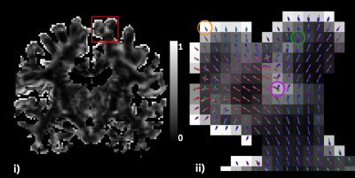 |
Investigating the Occurrence of Asymmetric Patterns in White Matter Fiber Orientation Distribution Functions
Charles Poirier1, Étienne St-Onge1, and Maxime Descoteaux1
1Sherbrooke Connectivity Imaging Laboratory (SCIL), Université de Sherbrooke, Sherbrooke, QC, Canada In general, fiber orientation distribution functions are defined as symmetric spherical functions. The present work shows how local averaging can be used to compute averaged asymmetric fODF (ava-fODF) from input symmetric white matter (WM) fODF. The resulting ava-fODF shows the presence of asymmetric patterns in voxels containing multiple crossings as well as along the white matter/gray matter interface. Moreover, the proportion of WM voxels displaying asymmetric features after filtering accounts for at least 30% of all WM voxels. Ava-fODF reveals new possibilities for classification of fiber configurations, with unidirectional fODF, bending fODF, Y-branching fODF and other complex fiber configurations. |
||
0866.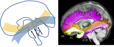 |
Beyond crossing fibers: investigating the prevalence of bottleneck configurations in the human brain white matter with diffusion tractography
Kurt G Schilling1, Francois Rheault2, Laurent Petit3, Chantal Tax4, Maxime Descoteaux2, Adam W Anderson5, and Bennett A Landman5
1Vanderbilt University Medical Center, Nashville, TN, United States, 2Universite de Sherbrooke, Sherbrooke, QC, Canada, 3CEA University of Bordeaux, Bordeaux, France, 4Cardiff University, Cardiff, United Kingdom, 5Vanderbilt University, Nashville, TN, United States
Bottleneck regions of the brain, or areas where multiple white matter pathways of the brain converge and subsequently diverge, present a challenge to anatomically accurate fiber tractography of the brain. In this work, we investigated the prevalence and locations of bottleneck regions. Our results indicate that most white matter contains multiple overlapping and crossing bundles. Moreover, individual orientations withina voxel are associated with multiple bundles, which represent bottlenecks. These findings have profound implications for tractography algorithms which aim to map unknown connections across the brain, and strengthen the awareness of limitations or challenges facing these image processing techniques.
|
||
|
0867.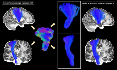 |
Bundle-o-graphy
Matteo Battocchio1, Simona Schiavi1, Maxime Descoteaux2, and Alessandro Daducci1
1University of Verona, Verona, Italy, 2University of Sherbrooke, Sherbrooke, QC, Canada
Here we introduce the concept of bundle-o-graphy to tackle tractography reconstruction from a different point of view: instead of indirectly reconstructing fiber bundles streamline-by-streamline, we consider them as the objects to be reconstructed and we directly seek for them using global optimization. We show the potential of bundle-based tractography on synthetic and real data, possibly opening avenues to a new way of thinking about tractography.
|
|
|
0868.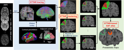 |
Atlas of reproducible short-range association fibers in parietal lobe by STTAR tracing and clustering
Chenying Zhao1,2, Minhui Ouyang1, and Hao Huang1,3
1Department of Radiology, Children's Hospital of Philadelphia, Philadelphia, PA, United States, 2Department of Bioengineering, School of Engineering and Applied Science, University of Pennsylvania, Philadelphia, PA, United States, 3Department of Radiology, Perelman School of Medicine, University of Pennsylvania, Philadelphia, PA, United States
Short-range association fibers (SAFs) linking adjacent cortices, are dominant in connectome and altered in psychiatric disorders such as autism and schizophrenia. However, few atlases are dedicated to SAFs. Higher inter-subject variation and large amount of SAFs impede their identification with traditional methods for long-range fibers. To meet these challenges, we built the SAF atlas using STTAR (Short-range Tractography with high Throughput And Reproducibility), a novel and advanced protocol designed for SAFs. Here, we demonstrated the SAF atlas in parietal lobe, featured with reproducibly and comprehensively traced SAFs readily usable in 3D volume format in the ICBM152 space.
|
The International Society for Magnetic Resonance in Medicine is accredited by the Accreditation Council for Continuing Medical Education to provide continuing medical education for physicians.
 Watch the Video