Digital Posters
Liver: Diffusion & Function
ISMRM & SMRT Annual Meeting • 15-20 May 2021

| Concurrent 5 | 15:00 - 16:00 |
2122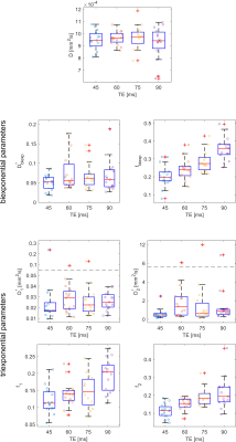 |
Echo time dependence of the biexponential and triexponential IVIM parameters in the liver Video Permission Withheld
Tobit Führes1, Andreas Julian Riexinger1, Jan Martin2, Martin Gerhard Loh1, Bernhard Hensel3, Michael Uder1, and Frederik Bernd Laun1
1University Hospital Erlangen, Erlangen, Germany, 2Lund University, Lund, Sweden, 3Friedrich-Alexander-Universität Erlangen-Nürnberg, Erlangen, Germany
In this study, we acquired diffusion-weighted image data of 15 healthy volunteers at four different TE. A ROI-based IVIM analysis showed that the pseudodiffusion coefficients did not change with TE, whereas the perfusion fractions rose significantly for bi- and triexponential IVIM curves at longer TE. A quantitative analysis indicates that the triexponential perfusion fractions are unlikely attributable to venous and arterial blood compartments. Our results indicate moreover that the pseudodiffusion coefficients can be compared without TE-correction among studies performed with different TE.
|
|||
2123.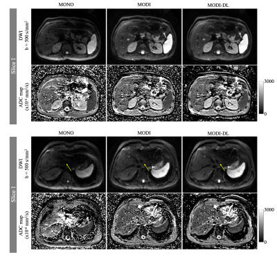 |
Motion-Robust, High-SNR Diffusion MRI of the Liver using Optimized Gradient Waveforms and Deep Learning Reconstruction
Yuxin Zhang1,2, Arnaud Guidon3, Xiaoili Zhao4, Gaohong Wu4, Hua Li4, Ruiqi Geng1,2, and Diego Hernando1,2
1Medical Physics, University of Wisconsin Madison, Madison, WI, United States, 2Radiology, University of Wisconsin Madison, Madison, WI, United States, 3GE Healthcare, Boston, MA, United States, 4GE Healthcare, Waukesha, WI, United States
Diffusion MRI of the liver has important applications in the detection of metastatic lesions. Unfortunately, conventional diffusion MRI is highly sensitive to cardiac-induced motion in the left lobe, whereas recently developed motion-robust diffusion MRI methods lead to lower SNR. This work demonstrates the feasibility of combining motion-robust diffusion MRI with DL reconstruction for improved DWI of the liver, including high motion robustness and high SNR. In early-stage evaluation, the combined technique provides excellent motion robustness, quantitative reliability, and high SNR throughout the entire liver, and may have application in the detection and evaluation of metastatic liver lesions.
|
|||
2124.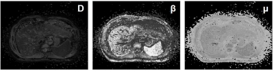 |
Staging chronic hepatitis B related liver fibrosis with a fractional order calculus diffusion model
Ruofan Sheng1, Yunfei Zhang2, Mengsu Zeng1, and Yongming Dai2
1Zhongshan Hospital affiliated to Fudan University, Shanghai, China, 2Central Research Institute, United Imaging Healthcare, Shanghai, China
Recently, a novel non-Gaussian DWI model, known as the fractional order calculus (FROC) model, was developed for biomedical applications. Until now, hardly has the FROC-DWI been applied in the hepatic imaging Thus, this study aimed to evaluate the value of FROC diffusion model in staging liver fibrosis. According to our results, the FROC-DWI-derived parameters (D, β, and μ) were significantly correlated with fibrosis stages, contributing positively to non-invasive assessment of liver fibrosis. The combination of FROC-DWI and conventional mono-exponential DWI held great potential in noninvasively staging liver fibrosis, superior to serum fibrosis biomarkers including APRI and FIB-4.
|
|||
2125.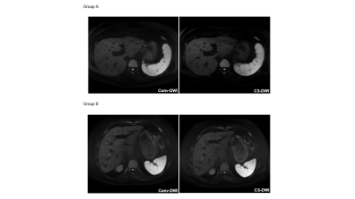 |
Liver Diffusion-Weighted MR Imaging with Compressed SENSE based on Single-Shot Echo-Planar Imaging: An intra-individual comparison
Maike Bode1, Shuo Zhang1,2, Nils A. Krämer1, Christiane K. Kuhl1, and Alexandra Barabasch1
1Diagnostic and Interventional Radiology, University Hospital RWTH Aachen, Aachen, Germany, 2Philips Healthcare, Hamburg, Germany
Compressed sensing (CS) has been reported to accelerate acquisition time in morphological imaging without image quality loss. The impact of CS on diffusion weighted imaging (DWI) in liver MRI has not yet been investigated. Therefore, the aim of our study was to test whether CS-based reconstruction is useful in echo-planar imaging (EPI) DWI. In this intra-individual comparison, EPI-DWI with CS-based reconstruction (CS-DWI) received comparable image quality ratings as conventional EPI-DWI. However, in a direct head-to-head-comparison, we found that a small fraction of focal liver lesions was missed on CS-DWI without apparent reason.
|
|||
2126.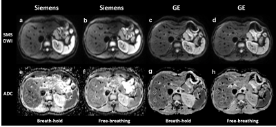 |
Reproducibility and Variability of Liver ADC Using Simultaneous Multi-slice DWI with Different Breathing Schemes and Different MR Vendors
Zheng Ye1, Bin Song1, Yuming Li1, Qing Li2, Lisha Nie3, and Xiaocheng Wei3
1West China Hospital, Sichuan University, Chengdu, China, 2MR collaborations, Siemens Healthcare Ltd., Shanghai, China, 3MR Research, GE Healthcare, Beijing, China
Diffusion-weighted imaging (DWI) provides anatomic and functional information, and apparent diffusion coefficient (ADC) has been proposed as a valuable biomarker in liver diseases. Simultaneous multi-slice (SMS) technique allows for exciting and acquiring multiple slices at the same time, and thus reducing scan time, which has been recently proved to be feasible in liver imaging. Our study investigated the reproducibility and variability of liver ADC of SMS-DWI with different breathing schemes and different vendors. We found good reproducibility of liver ADC across different MR vendors in both breath-hold and free-breathing manner. However, liver ADC is less reproducible between different breathing schemes.
|
|||
2127.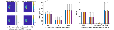 |
Optimization of b-values sampling scheme for several diffusion-weighted MRI models in the liver
Jiqing Huang1, Benjamin Leporq1, BEUF Olivier1, and Hélène Ratiney1
1Univ Lyon, INSA Lyon, CNRS, Inserm, CREATIS UMR 5220, U1206, F-69621, Lyon, Villeurbanne, France
To determine an optimal b-values sampling scheme for different non-Gaussian diffusion models in the liver, including diffusion kurtosis imaging (DKI), stretched-exponential model (SEM), intravoxel incoherent motion (IVIM), we optimized diffusion-weighting b-values sets using simulations mimicking real data and a b-value selection based on a Monte Carlo-like approach. Estimation performances were evaluated in terms of mean square error on signal and mean absolute percentage error on parameters in simulated data. The parameter estimation, with optimized set of b-values was finally applied on real data. The results showed that comparable fitting parameters and reconstructing signal can be obtained with fractional b-values.
|
|||
2128.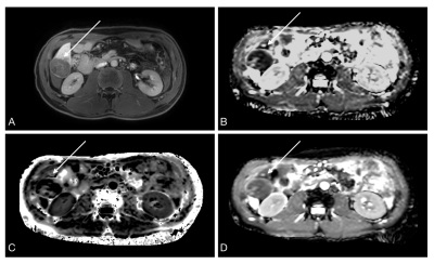 |
Diffusion kurtosis imaging (DKI) for predicting Ki-67 expression in hepatocellular carcinoma
Qiying Ke1, Tianyuan Zhang2, Xuan Jin1, Xinming Li1, Yingjie Mei3, and Xianyue Quan1
1Department of Radiology, Zhujiang Hospital, Southern Medical University, Guangzhou, China, 2Department of Radiology, Foshan First People's Hospital, Foshan, China, 3Philips Healthcare, Guangzhou, China
Diffusion kurtosis imaging (DKI) is a promising non-invasive method for evaluating the expression of Ki-67, a marker of cellular proliferation in HCC, which may contribute to predict recurrence and prognosis preoperatively. In this study, we evaluated the potential role of DKI in preoperative prediction of Ki-67 expression in HCC. The results show that increased MK and decreased ADC values are potential predictive biomarkers for of Ki-67 expression of in HCC.
|
|||
2129.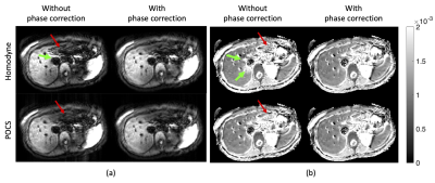 |
Phase correction in liver single-shot DW-EPI acquired with partial Fourier encoding
Anh T. Van1, Sean McTavish1, Johannes M. Peeters2, Kilian Weiss3, Marcus R. Makowski1, Rickmer F. Braren1, and Dimitrios C. Karampinos1
1Department of Diagnostic and Interventional Radiology, School of Medicine, Technical University of Munich, Munich, Germany, 2Philips Healthcare, Best, Netherlands, 3Philips Healthcare, Hamburg, Germany
In single shot diffusion imaging of moving organs, undesired motion during diffusion encoding introduces rapid phase variations in the obtained images. When partial Fourier encoding is used to reduce SNR loss in short T2 tissues, the motion-induced rapid phase variation compromises the partial Fourier reconstruction, resulting in “worm-like” artifacts in the reconstructed images. This work shows that by applying phase correction before the partial Fourier reconstruction motion-induced artifacts can be mitigated, improving the quality of diffusion-weighted images in liver applications.
|
|||
2130.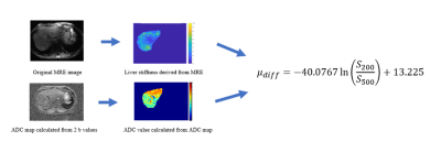 |
A New Diffusion-based Elastography for Quantitatively Assessing Liver Fibrosis without External Devices in a Large Set of Patients
Jie Yuan1, Fan Mo2, Yongming Dai2, Yuan Feng3, Suhao Qiu3, Songhua Zhan1, and Zhigang Gong1
1Department of Radiology, Shuguang Hospital Affiliated to Shanghai University of Traditional Chinese Medicine, 201203, Shanghai, China, 2MR Collaboration, United Imaging Healthcare, Shanghai, China, 3Institute for Medical Imaging Technology, School of Biomedical Engineering, Shanghai Jiao Tong University, 200240, Shanghai, China
Due to the non-invasive superiority, Magnetic Resonance Elastography (MRE) has emerged as an alternative approach to liver biopsy, the current golden standard for staging liver fibrosis. However, the use of external vibration devices brings costs of scan time and money. Diffusion Weighted Imaging (DWI) has shown the potential to characterize liver fibrosis at a microscopic level recently. The purpose of this study is to evaluate the potential of DWI to provide quantitative estimates of tissue stiffness without external devices among patients with different types of liver fibrosis and compare the inter-method agreement between MRE and the proposed method.
|
|||
2131.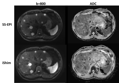 |
Comparison of integrated-shimming EPI and conventional SS-EPI diffusion-weighted MR imaging in Liver lesions
Hu Guo1, Huiting Zhang2, Junjiao Hu1, and Jun Liu1
1Department of Radiology, the Second Xiangya Hospital of Central South University, Changsha, China, 2MR Scientific Marketing, Siemens Healthcare, Wuhan, China The purpose of this study is to compare image quality and apparent diffusion coefficient (ADC) value between single-shot spin-echo echo-planar imaging (SS-EPI) and integrated slice-specific dynamic shimming (iShim) DWI methods in liver. Compared to SS-EPI, iShim had better image quality and showed more small lesions. No significant differences of ADC values were found between the two methods. |
|||
2132.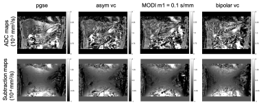 |
Gradient non-linearity correction in liver DWI using motion compensated diffusion encoding waveforms
Sean McTavish1, Anh T. Van1, Johannes M. Peeters2, Kilian Weiss3, Marcus R. Makowski1, Rickmer F. Braren1, and Dimitrios C. Karampinos1
1Department of Diagnostic and Interventional Radiology, Technical University of Munich, Munich, Germany, 2Philips Healthcare, Best, Netherlands, 3Philips Healthcare, Hamburg, Germany
Despite its strong clinical significance in lesion detection and tumor staging, liver DWI remains challenged by its strong sensitivity to motion effects. Motion-compensated diffusion encoding schemes have been recently proposed to improve DW liver signal homogeneity especially in the left liver lobe, a region typically affected by cardiac motion. In addition, a simplified gradient non-linearity correction scheme has been proposed for usage with standard pulsed gradient spin echo (pgse) diffusion encoding waveforms. The present work investigates the usage of this gradient nonlinearity correction algorithm with different motion compensated diffusion encoding gradient waveforms in the context of coronal ADC mapping in the liver.
|
|||
2133.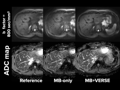 |
MultiBand-SENSE EPI with Variable-Rate Selective Excitation (VERSE) pulses for accelerating abdominal DWI with respiratory triggering
Kosuke Morita1, Masami Yoneyama2, Hiroshi Hamano2, Takeshi Nakaura3, Seitaro Oda3, Akira Sasao3, Hiroyuki Uetani3, Shogo Fukuda3, Masahiro Hatemura3, and Toshinori Hirai3
1Radiology, Kumamoto university, kumamoto-shi, Japan, 2Philips Japan, shinagawa, Japan, 3Kumamoto university, kumamoto-shi, Japan
We used respiratory triggered MultiBand SENSE (MB-SENSE) DWI using variable-rate selective excitation (MB-VERSE). We first measured ADC values of liver and kidney and subsequently performed visual assessment. The MB-VERSE DWI reduced the imaging time while improving the image quality and robustness of ADC values.
|
|||
2134.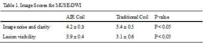 |
Comparison of Air Coil and the traditional abdomen coil for multiplexed sensitivity encoding diffusion weighted imaging (MUSE-DWI) of the liver
Wen Huiquan1, Zhang Yao2, Lin Wusheng2, Pi Shan2, Fang Ling3, and Wang Jin2
1Radiology, the Third Affiliated Hospital of Sun Yat-sen University (SYSU), Guangzhou, China, 2the Third Affiliated Hospital of Sun Yat-sen University (SYSU), Guangzhou, China, 3Department of Radiology, the Third Affiliated Hospital of Sun Yat-sen University (SYSU), Guangzhou, China
DWI is instrumental to screening and differential diagnosis of liver lesions. However, the diagnostic value DWI is often hindered by low SNR and severe image distortions, particularly in the left liver lobe. We explored the feasibility and efficacy of the combined use of AIR coil and multiplexed sensitivity encoding DWI (MUSE-DWI) in improving the image quality of liver. The results showed that MUSE-DWI with AIR Coil can effectively improve the image quality and reduce the image distortion of liver images, compared with the traditional abdomen coil, thus it is a feasible and effective method for detection of liver lesions.
|
|||
2135.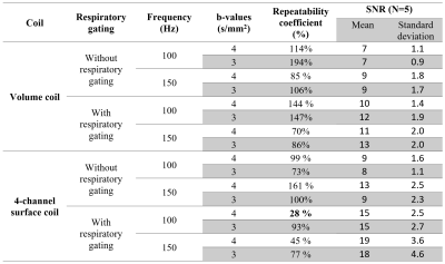 |
Temporal diffusion spectroscopy with oscillating gradient echoplanar MRI : signal to noise and ADC repeatability in mice liver.
Meryem Khalfallah1, Gwenaël Pagé1, Sabrina Doblas1, Bernard Van-Beers1,2, and Philippe Garteiser1
1Center of research on inflammation (UMR1149), Inserm - Université de Paris, Paris, France, 2Beaujon hospital radiology departement, APHP, Paris, France
Temporal diffusion spectroscopy using oscillating encoding gradients enables assessing tissue microstructure at a user-selected, arbitrary spatial scale. It has been demonstrated that this method is feasible with OGSE-EPI sequence. To study its robustness an OGSE-EPI sequence was implemented and repeatability and signal to noise ratio were assessed with two radiofrequency hardware setups and with or without respiratory gating. The effect of OGSE frequency was assessed for the pure diffusion coefficient (D) and for the apparent diffusion coefficient (ADC). Using a 4-channel surface coil and respiratory gating when measuring ADC at 100 Hz, the optimal repeatability coefficient (28%) was obtained.
|
|||
2136.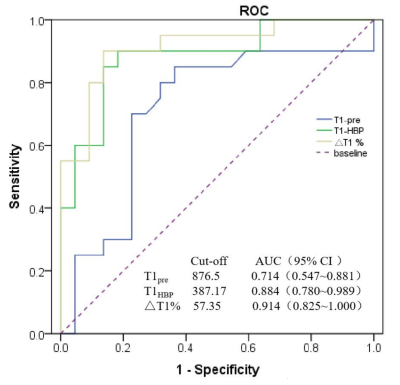 |
The diagnostic performance of Gd-EOB-DTPA-enhanced MRI based T1HBP value for liver function assessment
Pin Yang1, YanLi Jiang1, PengFei Wang1, TieJun Gan1, Rui Wang1, Jing Zhang1, and Kai Ai2
1Department of Magnetic Resonance, LanZhou University Second Hospital, Lanzhou, China, 2Philips Healthcare, Xi’an, China
The correlation between T1 relaxation times of hepatobiliary phase (T1HBP) and indocyanine green retention at 15min (ICG R15), as well as transient elastrography (TE) still need to be confirmed. This study evaluates the correlation among T1HBP, ICG R15 and TE. Our research showed that T1HBP had significant correlation with ICG R15 and TE. The above results demonstrated that T1HBP may be a promising radiography biomarker for liver function assessment.
|
|||
2137.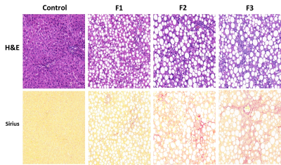 |
Detection of liver fibrosis in a Rat NASH Model using water specific T1 mapping with Gadoxetic Acid -enhanced MRI
Qian Wan1,2, Hao Peng 1, Jianxun Lyu1,2, Xiaoyi Liu3, Chuanli Cheng1, Feng Liu3, Yangzi Qiao1, Hairong Zheng1, Xin Liu1, Yi Wang3, and Chao Zou1
1Shenzhen Institutes of Advanced Technology, Chinese Academy of Sciences, Shenzhen, China, 2Shenzhen College of Advanced Technology,University of Chinese Academy of Sciences, Shenzhen, China, 3Peking University People's Hospital, Beijing, China
The severity of live fibrosis is one of the most important prognostic factors for the long-term outcomes of Nonalcoholic steatohepatitis (NASH). Early and precise diagnosis is of crucial importance for the disease management of NASH patients. In this work, we detected the liver fibrosis using water specific T1 mapping with Gadoxetic Acid -enhanced MRI in a rat NASH model induced by methionine-choline deficient (MCD) diet. The study revealed that the reduction rates of water-T1 relaxation time before and after Gd-EOB-DTPA administration might be a useful tool to stage the mild and moderate live fibrosis in rat NASH model.
|
|||
2138.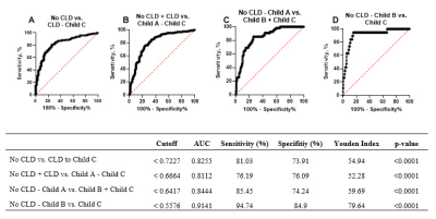 |
T1 reduction rate with Gd-EOB-DTPA to determine liver function in MRI – comparison between 1.5T and 3T in a cross-sectional study
Verena C. Obmann1, Damiano Catucci1, Annalisa Berzigotti2, Christoph Gräni3, Lukas Ebner1, Johannes Thomas Heverhagen1, Andreas Christe1, and Adrian Thomas Huber1
1Radiology, Inselspital, University of Bern, Bern, Switzerland, 2Visceral Surgery and Medicine, University of Bern, Inselspital, Bern, Switzerland, 3Cardiology, University of Bern, Inselspital, Bern, Switzerland
This study demonstrates that the MRI T1 reduction rate, based on T1 mapping before and after GD-EOB-DTPA, allows an accurate analysis of liver function and may be used interchangeably at 1.5T and 3T without image post-processing. T1 reduction rate was able to distinguish patients with no liver disease, patients with CLD and patients with liver cirrhosis Child A-C from one another.
|
|||
2139.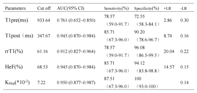 |
Quantitative Assessment of Liver Function by using Hepatocyte Fraction based on Gd-EOB-DTPA-Enhanced MRI
Xueqin ZHANG1, Jian LU1, Jifeng JIANG1, and Weibo CHEN2
1the Third People’s Hospital of Nantong, Nantong, China, 2Philips Healthcare, Shanghai, China
The purpose of this study was to assess the ability of hepatocyte fraction on Gd-EOB-DTPA-enhanced MRI for the quantitative evaluation of liver function. We used Look-Locker sequences to acquire T1 mapping images pre and post-contrast at 20 minutes after Gd-EOB-DTPA administration. T1 relaxation times (T1pre and T1post) of the liver, reduction rates of T1 relaxation times (rrT1), hepatocyte fraction (HeF) and KHep were measured. Our study showed that T1post, rrT1, HeF and KHep vlaues based on Gd-EOB-DTPA-enhanced T1 mapping MRI are efficient diagnostic tools for the quantitative evaluation of liver function.
|
|||
2140.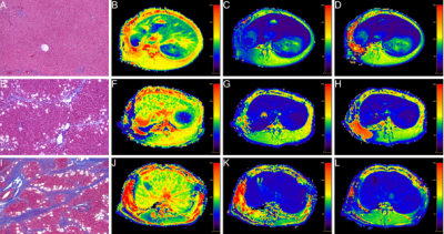 |
Study of Gd-EOB-DTPA T1mapping in quantitatively staging hepatic fibrosis in a rabbit model
Zuhui Zhu1, Wei Xing1, Haifeng Liu1, Qing Wang1, Yanan Du1, Yufeng Li1, and Jilei Zhang2
1Department of Radiology, third Affiliated Hospital of Soochow University & Changzhou First People's Hospital, Changzhou, China, 2Philips Healthcare, Shanghai, China To explore parameters obtained by Gd-EOB-DTPA T1 mapping and extracellular volume fraction in evaluating hepatic fibrosis (HF) in a rabbit model. The HF model was established by carbon tetrachloride (CCl4).Rabbits underwent pathological examination to determine the HF stage using the metavir classification system. Parameters including T1native, T110 min, T120 min, ECV10 min and ECV20 min were measured and compared among the different HF stages using spearman correlation coefficients and receiver operating characteristic curve. The AUC of T120 min was higher than that of other quantitative parameters. T1 mapping parameters and ECV fraction are closely associated with HF progression. |
|||
2141.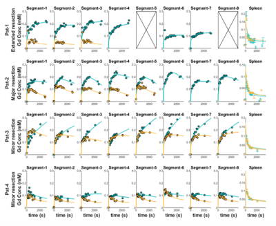 |
Regional Liver Function Pre- and Post-Resective Liver Surgery in Patients
Christian Simonsson1,2, Markus Karlsson1, Nils Dahlström1, Peter Lundberg1,2, Wolf Claus Bartholomä1,2, Gunnar Cedersund2,3, Per Sandström4, and Anna Lindhoff Larsson34
1Department of Radiation Physics, Radiology, Linköping University, Linköping, Sweden, 2Center for Medical Image Science and Visualization (CMIV), Linköping University, Linköping, Sweden, 3Department of Medical Engineering, Linköping University, Linköping, Sweden, 43Department of Surgery, Department of biomedical and clinical sciences, Linköping University, Linköping, Sweden
A range of severe liver diseases are identified only in very late stages. At a late stage, the only remaining treatment may be resective liver surgery. The resection can lead to serious complications if the remnant tissue fails to match the requirement of liver function. Therefore, it is valuable to accurately measure both regional and global liver function to better predict the outcome of the surgery. Here we use DCE-MRI to show that there are differences in Gadoxetate uptake both between patients and regional difference between the adjacent segments before and after resective surgery.
|
The International Society for Magnetic Resonance in Medicine is accredited by the Accreditation Council for Continuing Medical Education to provide continuing medical education for physicians.