Digital Posters
Breast: Breast Cancer Diagnosis, Characterization & Treatment Response
ISMRM & SMRT Annual Meeting • 15-20 May 2021

Digital Posters
Breast: Breast Cancer Diagnosis, Characterization & Treatment Response
| Concurrent 3 | 17:00 - 18:00 |
1431.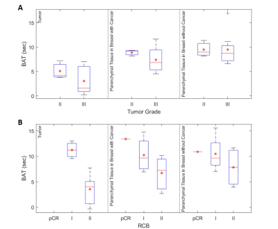 |
Kinetic parameters derived from ultrafast DCE-MRI to predict breast cancer response to neoadjuvant chemotherapy
Zhen Ren1, Federico D Pineda1, Elle Hill1, Teodora Szasz1, Rabia Safi1, Chengyue Wu2, Thomas E Yankeelov2, Kirti Kulkarni1, Hiroyuki Abe1, Rita Nanda1, and Gregory S Karczmar1
1University of Chicago, Chicago, IL, United States, 2The University of Texas at Austin, Austin, TX, United States
We retrospectively reviewed data from 11 patients who received NAC and were scanned with a protocol that included ultrafast DCE-MRI for the first minute after contrast injection, followed by high spatial-resolution post-contrast imaging prior to therapy and post-treatment. The results showed a significant correlation between response to therapy and the contrast media bolus arrival time (BAT) in tumor, and normal parenchyma in each breast separately. In addition, BAT is significantly associated with background parenchymal enhancement (BPE) while BPE rate is independent of BPE. BPE rate and tumor enhancement rate may be independent predictors for malignancy.
|
|||
1432.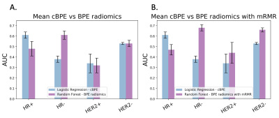 |
Background Parenchymal Enhancement Radiomic Features for Neoadjuvant Treatment Response Prediction in Breast Cancer Patients
Alex Anh-Tu Nguyen1, Natsuko Onishi1, Wen Li1, Deep K Hathi1, Rohan Nadkarni1, Efstathios D Gennatas2, Ella F Jones1, I-SPY2 Consortium3, David C Newitt1, and Nola M Hylton1
1Radiology & Biomedical Imaging, University of California, San Francisco, San Francisco, CA, United States, 2Epidemiology & Biostatistics, University of California, San Francisco, San Francisco, CA, United States, 3Quantum Leap Healthcare Collaborative, San Francisco, CA, United States
Background parenchymal enhancement (BPE) observed in normal fibroglandular tissue in breast dynamic contrast-enhanced MRI shows an association with breast cancer risk and has been investigated to predict treatment response. Contralateral BPE radiomic features and mean BPE values were calculated from DCE-MRI and their predictive performance of pathologic complete response after neoadjuvant chemotherapy was tested. The best mean AUCs were found using BPE radiomics based random forest model with minimal-redundancy-maximal-relevance feature selection in the HR- group and HER2- group. Our results show that contralateral BPE radiomic features have potential as imaging biomarkers for treatment response.
|
|||
1433. |
Predictive value of APTw for early evaluation of pathological complete response to NAC in molecular subtyping of breast cancer
Nan Zhang1, Lina Zhang1, Qingwei Song1, Ailian Liu1, Keshuo Tang2, Lei Dong2, Jiazheng Wang3, and Zhiwei Shen3
1The First Affiliated Hospital of Dalian Medical University, Dalian, China, 2Dalian Medical University, Dalian, China, 3Philips Healthcare, Beijing, China, Beijing, China
Amide proton transfer (APT) imaging is based on the chemical exchange between free bulk water protons and the amide protons (-NH) of endogenous mobile proteins and peptides in tissue[1]. Nan Meng et al suggested that APTw imaging can be used for the differential diagnosis of benign and malignant breast lesions[2]. A simple sample study has shown that APTw MRI provides a possible biomarker for assessing chemotherapy response in human breast cancer patients on 3T MRI[3]. This study aims to explore the feasibility of APTw-MRI in early evaluation of pathological complete response to NAC in molecular subtyping of breast cancer
|
|||
1434.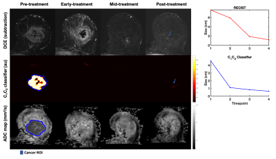 |
Three-component diffusion model with deformable registration for automated evaluation of response to neoadjuvant therapy in breast cancer
Maren M. Sjaastad Andreassen1, Michelle W. Tong2, Ana E. Rodríguez-Soto2, Somaye Zare3, Tyler M. Seibert2,4,5, Michael Hahn2, Haydee Ojeda-Fournier 2, Neil P. Jerome1,6, Tone F. Bathen1,7, Anders M. Dale2,8, and Rebecca Rakow-Penner2
1Department of Circulation and Medical Imaging, Norwegian University of Science and Technology, Trondheim, Norway, 2Department of Radiology, University of California San Diego, La Jolla, CA, United States, 3Department of Pathology, University of California San Diego, La Jolla, CA, United States, 4Department of Bioengineering, University of California San Diego, La Jolla, CA, United States, 5Department of Radiation Medicine and Applied Sciences, University of California San Diego, La Jolla, CA, United States, 6Department of Physics, Norwegian University of Science and Technology, Trondheim, Norway, 7Department of Radiology and Nuclear Medicine, Norwegian University of Science and Technology, Trondheim, Norway, 8Department of Neuroscience, University of California San Diego, La Jolla, CA, United States
The purpose of this work was to develop a method to automatically monitor response to neoadjuvant treatment in breast cancer using longitudinally registered multi-b diffusion MRI acquisitions. The two slowest diffusion signal contributions from a three-component model were used to generate cancer probability maps (C1C2 classifier) that estimated tumor volume at each timepoint. Our results demonstrated that changes in C1C2 classifier agreed with standard dynamic-contrast-enhanced MRI criteria (RECIST) in 23 out of 24 cases. In 35% of treatment responders, the C1C2 classifier captured response at an earlier timepoint than RECIST.
|
|||
1435.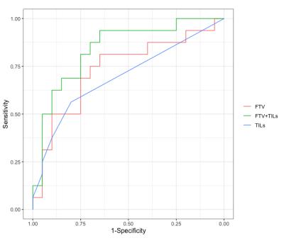 |
Associations of pretreatment MRI with pathologic assessment of immune infiltration and chemotherapy response in triple negative breast cancer
Anum S. Kazerouni1, Laura C. Kennedy2, Michael Hirano1, Daniel S. Hippe1, Bonny Chau1, Debosmita Biswas1, Wesley Surento1, Shaveta Vinayak1, Matthew Nyflot1, Habib Rahbar1, Suzanne Dintzis1, and Savannah C. Partridge1
1University of Washington, Seattle, WA, United States, 2Vanderbilt University, Nashville, TN, United States
We investigated the relationship between immune infiltration and imaging metrics derived from breast MRI in patients with triple negative breast cancer (TNBC) treated with neoadjuvant chemotherapy (NAC). Thirty-six patients with localized TNBC were imaged with diffusion-weighted and dynamic contrast-enhanced MRI prior to initiation of NAC. Baseline tumor infiltrating lymphocytes (TILs) and functional tumor volume were found to significantly differ between patients who achieved pathological complete response (pCR) compared to non-pCR patients. Baseline ADC showed a weak negative correlation with TIL counts.
|
|||
1436.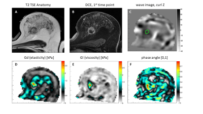 |
Measuring the tumour IFP using MRE: a mechanical biomarker for the prediction of metastatic potential in women with invasive breast cancer
Patriek Jurrius1,2, Omar Darwish1,3,4, Belul Shifa2, Joanna Bell2, John Spence2, Daniel Fovargue1, Giacomo Annio3, Renee Miller1, Marian Troelstra1, Ashutosh Kothari2, Hisham Hamed2, Diana Stavrou2, Ali Sever2, Sultana Hasso2,
Katerina Ntailiani2, Keshthra Satchithananda5, Sarah Willson2, Sarah Pinder1,2, Anne Rigg2, David Nordsletten1, Radhouene Neji1,4, Arnie Purushotham1,2, and Ralph Sinkus1,3
1King's College London, London, United Kingdom, 2Guy's and St. Thomas' NHS Foundation Trust, London, United Kingdom, 3University Paris Diderot, Paris, France, 4Siemens Healthcare Ltd., Frimley, United Kingdom, 5King's College Hospital NHS Foundation Trust, London, United Kingdom
Breast cancer is the most prevalent cancer among women. Over the years the overall survival has increased, with metastatic dissemination being one the most important prognostic factors. Currently, the presence of lymph node involvement or distant metastasis is diagnosed on imaging and core biopsy results. Yet, no technique to predict a cancer’s metastatic potential a priori is available. By incorporating magnetic resonance elastography (MRE) in a patient’s routine MRI scan the tumour interstitial fluid pressure, a factor associated with metastatic propensity, can be non-invasively measured. Preliminary results indicate a correlation between MRE measured tumour pressure and the cancer’s metastatic potential.
|
|||
1437.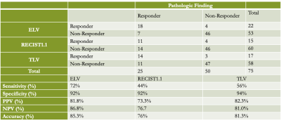 |
Feasibility of quantitative and volumetric enhancement measurement to assess tumor response in patients with breast cancer after early NAC
Jie Ding1, Ruoshui Ha1, Weiwei Deng2, and Xiuzheng Yue3
1Medical Imaging Center, People's Hospital of Ningxia Hui Autonomous Region, Yinchuan, China, 2Philips Healthcare, Shanghai, China, 3Philips Healthcare, Beijing, China The accuracy of early efficacy evaluation of neoadjuvant chemotherapy for breast cancer is important for clinicians to choose chemotherapy regimen. 75 lesions were assessed according to the ELV method in comparison with RECIST1.1 and TLV. In terms of predicting pathological response, the diagnostic accuracy of the three methods were analyzed and compared by receiver operating characteristic (ROC) curve analysis. It is feasible for ELV in evaluating the responsiveness of breast cancer patients to early NAC on DCE-MRI images. Referring to the prediction the pathological response, ELV had the best diagnostic performance with highest sensitivity, accuracy, as well as AUC value. |
|||
1438.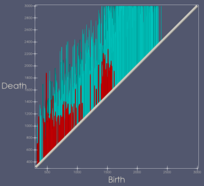 |
DCE-MRI Topology for Early Prediction of Breast Cancer Response to Neoadjuvant Chemotherapy
Josiah D Blaisdell1, Wei Huang2, and Yue Zhang1
1EECS, Oregon State University, Corvallis, OR, United States, 2Advanced Imaging Research Center, Oregon Health & Science University, Portland, OR, United States
We applied Topological Data Analysis (TDA) to pre-neoadjuvant chemotherapy (pre-NAC) DCE-MRI datasets from the ISPY-1 trial for NAC treatment of breast cancer (BC). The pairwise topological distance between tumors’ signal enhancement ratio (SER) maps was computed. Hierarchical Agglomerative Clustering (HAC) was applied to cluster topologically similar patients. In combination with clinical and histopathological data using logistic regression models, the predictive performance of MRI topology for pathologic complete response (pCR) was compared to longest diameter (LD) and functional tumor volume (FTV). The preliminary results show that MRI topology may be a more accurate early predictor of BC response to NAC.
|
|||
1439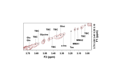 |
Tentative assignment of methylmalonic acid in the breast tissue using in vivo 2D COSY and relationships with risk factors of breast cancer Video Permission Withheld
Gorane Santamaría1,2, Natali Naude2,3, Peter Malycha3,4, and Carolyn Mountford2,3
1Radiology, Princess Alexandra Hospital, Brisbane, Australia, 2Queensland University of Technology, Brisbane, Australia, 3Translational Research Institute, Brisbane, Australia, 4Jones and Partners Radiology, St Andrew’s Hospital, Adelaide, Australia
Methylmalonic acid (MMA) has been linked to ageing and a systemic environment that favours the progression and tumours aggressiveness. We aim to assign MMA in breast tissue using in vivo 2D COSY in a series of 172 participants including women at average and elevated risk. A second purpose is to explore if MMA is associated with breast cancer risk factors. Two resonances were recorded at 3.15 and 3.19ppm in breast tissue, consistent with the presence of MMA1 and MMA2 molecules. The level of MMA correlates positively with breast density/menopausal status and IBIS risk score and, negatively with age and BMI.
|
|||
1440.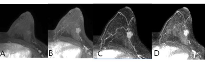 |
Ultrafast DCE MRI Using Compressed Sensing: Separate Visualization of Arteries and Veins in Differential Diagnosis of Breast Tumors (BI-RADS 4)
Nan Zhang1, Zhuo Wang2, Liguo Tan2, Weizheng Chen2, Lina Zhang1, Ailian Liu1, Qingwei Song1, Jiazheng Wang3, and Zhiwei Shen3
1The First Affiliated Hospital of Dalian Medical University, Dalian, China, 2Dalian Medical University, Dalian, China, 3Philips Healthcare, Beijing, China, Beijing, China
Dynamic contrast-enhanced magnetic resonance imaging (DCE-MRI) is an important tool for the diagnosis of breast cancer. It is of great importance to develop new MRI acquisition and analysis methods that can yield improved biomarkers with higher specificity for the diagnosis of breast lesions. The purpose of the present study is to explore visualization and quantitative information of tumor-associated vessels to improve diagnostic accuracy of breast tumors (MR BI-RADS 4). The results show that combination of quantitative breast vascular information may provide an accurate means for the diagnosis of breast cancer.
|
|||
1441.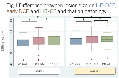 |
Can we use Ultrafast Dynamic Contrast Enhanced MRI to evaluate Ductal Carcinoma in situ?
Masako Y Kataoka1, Maya Honda1, Mami Iima1, Akane Ohashi2, Rie Ota1, Yosuke Yamada3, Masakazu Toi4, and Yuji Nakamoto1
1Diagnostic Imaging and Nuclear Medicine, Kyoto University Graduate School of Medicine, Kyoto, Japan, 2Kyoto Medical Center, Kyoto, Japan, 3Department of Diagnostic Pathology, Kyoto University Hospital, Kyoto, Japan, 4Department of Breast Surgery, Kyoto University Hospital, Kyoto, Japan
Using ultrafast DCE (UF-DCE) MRI, size and morphology of DCIS on very early phase of the post contrast injection were compared to those on early phase of conventional DCE (C-DCE) MRI and on High resolution post contrast enhancement (HR-CE). Lesion size tended to be smaller on UF-DCE MRI. Clustered ring is not frequently observed on UF-DCE MRI compared to HR-CE. These data suggest evaluating DCIS on UF-DCE may be dealt with caution.
|
|||
1442.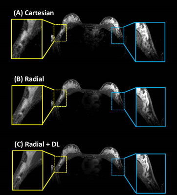 |
Deep learning reconstruction including de-streaking capability for motion-robust T1-weighted breast imaging
Ping N Wang1, Sagar Mandava2, Xinzeng Wang3, Ty A Cashen4, Frederick Felcz5, and James H Holmes5
1Department of Medical Physics, University of Wisconsin-Madison, Madison, WI, United States, 2Global MR Applications and Workflow, GE Healthcare, Atlanta, GA, United States, 3Global MR Applications and Workflow, GE Healthcare, Houston, TX, United States, 4Global MR Applications and Workflow, GE Healthcare, Madison, WI, United States, 5Department of Radiology, University of Wisconsin-Madison, Madison, WI, United States
DCE imaging is the primary technique for MR evaluation of breast cancer. A key problem is ghosting due to cardiac motion obscuring axillary breast tissue. Addressing this challenge, motion-insensitive technologies such as stack-of-stars acquisition have been proposed, which introduces its own problem of streaking. Based on success with a DL reconstruction to reduce noise, blurring, and ringing, this work investigated re-purposing this deep CNN to also mitigate streaking. Phantom imaging demonstrated improved CNR and more accurate line profiles. 15 patients undergoing a clinical MR exam were scanned with the additional motion-robust method, and images showed reduced structured/unstructured noise and blurring.
|
|||
1443.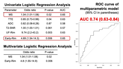 |
Multiparametric prediction model for triple negative breast cancer subtypes using MR parameters including ultrafast DCE MRI
Akane Ohashi1, Masako Kataoka2, Mami Iima2, Maya Honda2, Rie Ota2, Yuta Urushibata 3, Nickel Marcel Dominic 4, Masakazu Toi5, Yuusuke Hirokawa1, and Yuji Nakamoto2
1Radiology, National Hospital Organisation, Kyoto Medical Center, Kyoto, Japan, 2Diagnostic Imaging and Nuclear Medicine, Kyoto University Graduate School of Medicine, Kyoto, Japan, 3Siemens Healthcare K.K., Tokyo, Japan, 4Siemens Healthcare GmbH, Erlangen, Germany, 5Breast Surgery, Kyoto University Graduate School of Medicine, Kyoto, Japan
We tried to construct MRI-based multiparametric model to predict triple negative (TN) subtype among invasive cancers presenting as masses using 165 lesions (including 26 TN subtype). Maximum slope (MS) and time to enhancement (TTE) from ultrafast (UF)-DCE MRI, apparent diffusion coefficient (ADC), signal to noise ratio on T2-WI, rim enhancement on different phases of the DCE MRI were examined with univariate and multivariate logistic regression analysis. The model using MS from UF-DCE MRI and rim enhancement from early phase of DCE MRI demonstrated the AUC of 0.74 in identifying TN subtype, indicating the MRI’s potential to identify TN subtype noninvasively.
|
|||
1444.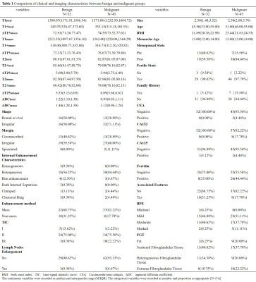 |
BI-RADS 4 Breast Lesions: Could Synthetic MRI Be Helpful for Their Diagnosis?
Shi Yun Sun1, Zhuo Lin Li1, Ying Ying Ding1, Li Sha Nie2, Cheng De Liao1, Yi Fan Liu1, Rui Wang1, Jia Zhang3, and Dong Xue Zhang1
1The Third Affiliated Hospital of Kunming Medical University,Yunnan Cancer Hospital, Kunming, China, 2GE Healthcare, MR Research China, Beijing, China, Beijing, China, 3The Third People's Hospital of Yunnan Province, China, Kunming, China
Synthetic MRI (syMRI) can quantify multiple relaxation parameters at the same time, which might have potential application value in the BI-RADS 4 lesions. 77 breast disease patients who were defined as BI-RADS 4 in the preoperative MRI examination were prospectively enrolled in this study. Before and after contrast injection, all patients underwent routine MRI and syMRI examinations. The result show that relaxation time and ADC values provided by syMRI and DWI are useful in distinguishing breast BI-RADS 4 lesions. The multi parameter model combined with clinical and imaging features can significantly improve the diagnostic ability of BI-RADS 4 breast lesions.
|
|||
1445.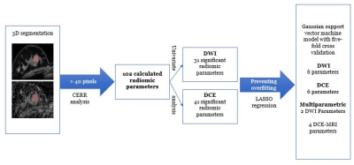 |
Radiomics and Machine Learning on multiparametric MRI for breast cancer diagnosis
Isaac Daimiel Naranjo1, Roberto Lo Gullo2, Carolina Sacarelli2, Almir Bitencourt2, Peter Gibbs2, Elisabeth Morris2, Caleb Sooknanan2, Jeff Reiner2, Maxine S Jochelson2, Sunitha Thakur2, and Katja Pinker-Domenig2
1Radiology, Memorial Sloan Kettering Cancer Center, NEW YORK, NY, United States, 2Memorial Sloan Kettering Cancer Center, New york, NY, United States
Radiomics coupled with machine learning is based on the extraction of signatures from medical images that are invisible to the human eye to create models which would improve breast cancer diagnosis. Radiomics features extracted from dynamic contrast-enhanced MRI and diffusion-weighted imaging can be combined in multiparametric MRI. We hypothesize that radiomics features extracted from multiparametric MRI would allow for an improved model affording a more accurate breast cancer diagnosis. We developed a multiparametric model that achieved the best accuracy for breast cancer diagnosis compared to models based on dynamic contrast-enhanced MRI or diffusion-weighted imaging.
|
|||
1446.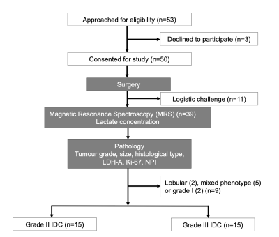 |
Lactate concentration in breast cancer using advanced magnetic resonance spectroscopy
Sai Man Cheung1, Ehab Husain2,3, Iain D Miller2, Yazan Masannat3,4, Klaus Wahle5, Steven D Heys4, and Jiabao He1
1Institute of Medical Sciences, University of Aberdeen, Aberdeen, United Kingdom, 2Pathology Department, Aberdeen Royal Infirmary, Aberdeen, United Kingdom, 3School of Medicine, University of Aberdeen, Aberdeen, United Kingdom, 4Breast Unit, Aberdeen Royal Infirmary, Aberdeen, United Kingdom, 5Strathclyde Institute of Pharmacy and Biological Sciences, University of Strathclyde, Glasgow, United Kingdom
Upregulation of aerobic glycolysis and an elevated lactate accumulation have been linked to tumour aggressiveness. However, current evidence drawn from cell culture and small animal models remains controversial. Since lactate and lipid share the same spectral frequency, conventional MRS is inadequate in quantification of lactate under overwhelming lipid signal. Double quantum filtered (DQF) MRS allows excellent suppression of lipid signal from adipose breast tissues. We conducted a cross sectional study to examine the prognostic role of lactate concentration in grade II and III whole breast tumours using DQF MRS for the quantification of lactate concentration.
|
|||
1447.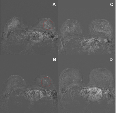 |
Systemic effects of intrauterine contraceptive devices demonstrated by background parenchymal enhancement in DCE-MRI.
Luisa Charlotte Huck1, Daniel Truhn2, Ebba Dethlefsen2, and Christiane Katharina Kuhl2
1Radiology, RWTH Aachen University, Aachen, Germany, 2RWTH Aachen University, Aachen, Germany According to the manufacturers intrauterine contraceptive devices (IUDs) should not be associated with systemic hormonal effects. However, women’s representatives claim that IUDs do exhibit side effects that are similar to oral or percutaneous hormonal medication. Background parenchymal enhancement (BPE) reduces the sensitivity and specificity of breast MRI and is influenced by exogenous hormonal exposures. We investigated whether IUD use is associated with BPE on contrast-enhanced breast MRI, as a surrogate marker for hormonal stimulation. We found that the application of an IUD is associated with increased BPE in breast MRI. This suggests that IUDs do have a systemic hormonal effect.
|
|||
1448.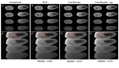 |
Implementing ConvDecoder with physics-based regularization to reconstruct under-sampled variable-flip angle MRI data of the breast
Kalina P Slavkova1, Julie C DiCarlo2,3, Viraj Wadhwa4, Jingfei Ma5, Gaiane M Rauch6, Zijian Zhou5, Thomas E Yankeelov2,3,7,8,9, and Jonathan I Tamir2,4,8
1Department of Physics, The University of Texas at Austin, Austin, TX, United States, 2Oden Institute for Computational Engineering and Sciences, The University of Texas at Austin, Austin, TX, United States, 3Livestrong Cancer Institutes, The University of Texas at Austin, Austin, TX, United States, 4Department of Electrical and Computer Engineering, The University of Texas at Austin, Austin, TX, United States, 5Department of Imaging Physics, MD Anderson Cancer Center, Houston, TX, United States, 6Department of Abdominal Imaging, MD Anderson Cancer Center, Houston, TX, United States, 7Department of Biomedical Engineering, The University of Texas at Austin, Austin, TX, United States, 8Department of Diagnostic Medicine, The University of Texas at Austin, Austin, TX, United States, 9Department of Oncology, The University of Texas at Austin, Austin, TX, United States
We evaluate the ability of the ConvDecoder architecture regularized using a physical model to reconstruct under-sampled dynamic MRI data, namely variable-flip angle data as a proof-of-principle. The performance of the reconstruction is evaluated by comparing the normalized error with results returned by compressed sensing and the non-regularized ConvDecoder. We hypothesize that ConvDecoder with physics-based regularization will enable significantly fewer k-space measurements, thereby allowing for expedited scan time while maintaining spatial resolution.
|
|||
1449.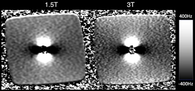 |
Systematic characterization of MRI near a surgical breast implant at 1.5T and 3.0T
Collin J Buelo1,2, Scott B Reeder1,2,3,4,5, and Diego Hernando1,2,5
1Medical Physics, University of Wisconsin-Madison, Madison, WI, United States, 2Radiology, University of Wisconsin-Madison, Madison, WI, United States, 3Emergency Medicine, University of Wisconsin-Madison, Madison, WI, United States, 4Medicine, University of Wisconsin-Madison, Madison, WI, United States, 5Biomedical Engineering, University of Wisconsin-Madison, Madison, WI, United States
Metallic clip soft tissue markers are a promising alternative to wire-guided localization in localizing breast cancer for surgery. However, the metallic core in the marker creates magnetic field distortions with MRI, which leads to various artifacts including signal voids and failed chemical shift-based fat suppression. This work characterizes the effect of a soft tissue marker at 1.5T and 3.0T using conventional breast imaging sequences as well as multi-spectral sequences. Several fat suppression methods were also evaluated. Field distortions and imaging artifacts are similar at 1.5T and 3.0T, and can be addressed using multi-spectral methods and T1 based fat suppression.
|
|||
1450.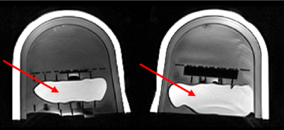 |
Initial Results of a Novel High-Resolution 3D-FSE Based Protocol for Silicone Breast Implant Screening
Tayeb Zaidi1, John Mugler2, Maureen Hood3, Brian Garra4, Erin Kelly5, Hung Do5, Mo Kadbi5, and Sunder Rajan1
1Division of Biomedical Physics, Food and Drug Administration, Silver Spring, MD, United States, 2Department of Radiology and Medical Imaging Research, University of Virginia, Charlottesville, VA, United States, 3Department of Radiology & Radiological Sciences, Uniformed Services University, Bethesda, MD, United States, 4Division of Imaging, Diagnostics, and Software Reliability, Food and Drug Administration, Silver Spring, MD, United States, 5Canon Medical Systems USA, Tustin, CA, United States
Conventional silicone breast MR involves multi-contrast 2D scans. However, the use of 2D imaging risks the loss of fine structure visualization in the 3rd dimension. An isotropic 3D-FSE pulse sequence was first tested in a single-center study in 13 subjects with silicone breast implants (SBI). The images were evaluated by radiologists for quality and added utility of the bilateral, 3D scans. To allow for multi-site implementation of the 3D sequence, a bilateral breast phantom containing lipid, fibroglandular mimic and a silicone implant was tested across major MR vendors to ensure adequate signal-to-noise ratios and tissue contrast.
|
The International Society for Magnetic Resonance in Medicine is accredited by the Accreditation Council for Continuing Medical Education to provide continuing medical education for physicians.